Abstract
The muscles of the upper extremity, comprising the shoulder, arm, forearm, and hand, are essential for a wide range of movements, from gross motor functions like lifting and pushing to fine motor skills such as writing and manipulating objects. These muscles are categorized based on their anatomical location and function. This article provides an in-depth examination of the anatomy, development, functions, and clinical significance of the muscles of the upper extremity.
Introduction
The upper extremity muscles play a crucial role in the versatility and dexterity of the human body. They enable a broad spectrum of movements necessary for daily activities, sports, and complex tasks. Understanding the anatomy and function of these muscles is vital in fields such as orthopedics, physical therapy, sports medicine, and neurology.
Anatomical Structure
The muscles of the upper extremity can be categorized into four main groups based on their location and function:
- Muscles of the Shoulder (Scapular and Thoracic Region)
- Muscles of the Arm (Brachium)
- Muscles of the Forearm (Antebrachium)
- Muscles of the Hand
Muscles of the Shoulder
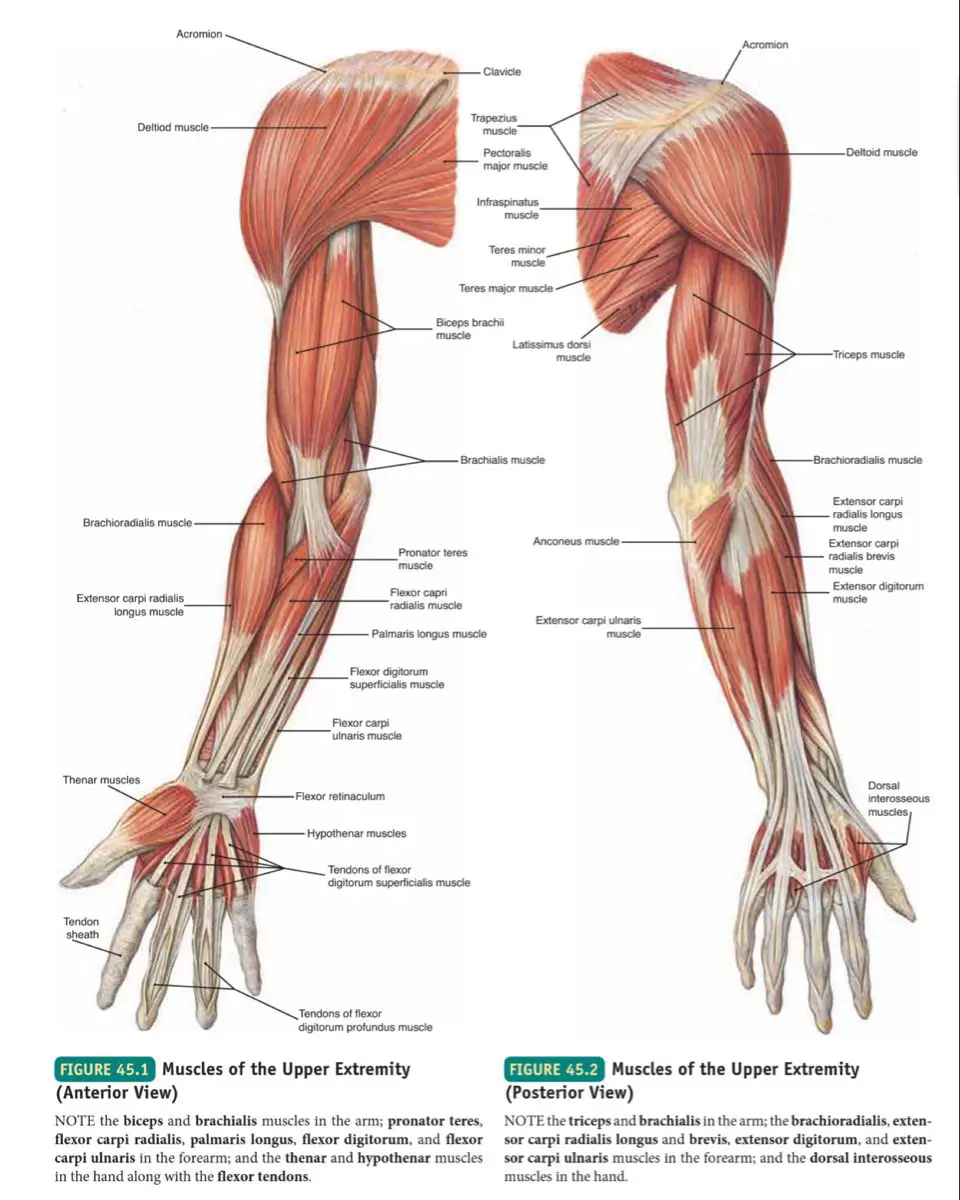
The shoulder muscles are primarily responsible for stabilizing the shoulder joint and facilitating arm movements. They can be divided into two groups: the extrinsic and intrinsic muscles.
Extrinsic Shoulder Muscles
These muscles originate on the torso and insert on the bones of the shoulder girdle.
- Trapezius: Elevates, retracts, and rotates the scapula; also involved in extending the neck.
- Latissimus Dorsi: Extends, adducts, and medially rotates the arm.
- Levator Scapulae: Elevates the scapula and assists in neck rotation.
- Rhomboid Major and Minor: Retract and rotate the scapula.
Intrinsic Shoulder Muscles
These muscles originate on the scapula and clavicle and insert on the humerus.
- Deltoid: Abducts, flexes, and extends the arm; responsible for the rounded contour of the shoulder.
- Teres Major: Assists in the adduction, extension, and medial rotation of the arm.
- Rotator Cuff Muscles: A group of four muscles that stabilize the shoulder joint:
- Supraspinatus: Abducts the arm.
- Infraspinatus: Externally rotates the arm.
- Teres Minor: Externally rotates and adducts the arm.
- Subscapularis: Internally rotates the arm.
Muscles of the Arm
The muscles of the arm are primarily involved in flexion and extension of the elbow and shoulder joints. They are divided into anterior (flexor) and posterior (extensor) compartments.
Anterior Compartment
- Biceps Brachii: A two-headed muscle that flexes the elbow and supinates the forearm; also assists in shoulder flexion.
- Brachialis: Lies deep to the biceps brachii and is the primary flexor of the elbow.
- Coracobrachialis: Flexes and adducts the arm at the shoulder joint.
Posterior Compartment
- Triceps Brachii: A three-headed muscle that extends the elbow; the long head also assists in shoulder extension and adduction.
- Anconeus: Assists the triceps brachii in extending the elbow and stabilizing the elbow joint.
Muscles of the Forearm
The forearm muscles control movements of the wrist, hand, and fingers. They are divided into anterior (flexor) and posterior (extensor) compartments.
Anterior Compartment
These muscles are primarily involved in flexion of the wrist and fingers, and pronation of the forearm. They are further divided into superficial, intermediate, and deep layers.
- Superficial Layer:
- Pronator Teres: Pronates the forearm.
- Flexor Carpi Radialis: Flexes and abducts the wrist.
- Palmaris Longus: Flexes the wrist and tenses the palmar fascia.
- Flexor Carpi Ulnaris: Flexes and adducts the wrist.
- Intermediate Layer:
- Flexor Digitorum Superficialis: Flexes the middle phalanges of the fingers.
- Deep Layer:
- Flexor Digitorum Profundus: Flexes the distal phalanges of the fingers.
- Flexor Pollicis Longus: Flexes the thumb.
- Pronator Quadratus: Pronates the forearm.
Posterior Compartment
These muscles are primarily involved in extension of the wrist and fingers, and supination of the forearm. They are also divided into superficial and deep layers.
- Superficial Layer:
- Brachioradialis: Flexes the forearm at the elbow, especially when the forearm is in a mid-position between pronation and supination.
- Extensor Carpi Radialis Longus: Extends and abducts the wrist.
- Extensor Carpi Radialis Brevis: Extends and abducts the wrist.
- Extensor Digitorum: Extends the fingers and the wrist.
- Extensor Digiti Minimi: Extends the little finger.
- Extensor Carpi Ulnaris: Extends and adducts the wrist.
- Deep Layer:
- Supinator: Supinates the forearm.
- Abductor Pollicis Longus: Abducts and extends the thumb.
- Extensor Pollicis Longus and Brevis: Extend the thumb.
- Extensor Indicis: Extends the index finger.
Muscles of the Hand
The muscles of the hand are responsible for fine motor movements, including gripping, grasping, and manipulating objects. They are divided into intrinsic and extrinsic muscles.
Intrinsic Muscles
These muscles are located entirely within the hand and are involved in precise finger movements.
- Thenar Muscles: Control the movements of the thumb.
- Abductor Pollicis Brevis: Abducts the thumb.
- Flexor Pollicis Brevis: Flexes the thumb.
- Opponens Pollicis: Opposes the thumb.
- Hypothenar Muscles: Control the movements of the little finger.
- Abductor Digiti Minimi: Abducts the little finger.
- Flexor Digiti Minimi Brevis: Flexes the little finger.
- Opponens Digiti Minimi: Opposes the little finger.
- Lumbricals: Flex the metacarpophalangeal joints and extend the interphalangeal joints of the fingers.
- Interossei Muscles: Consist of dorsal (which abduct the fingers) and palmar (which adduct the fingers) interossei muscles, important for finger movements and grip strength.
- Adductor Pollicis: Adducts the thumb.
Extrinsic Muscles
These muscles originate in the forearm and insert on the hand, controlling gross movements of the fingers and wrist.
- Flexor Digitorum Superficialis: Flexes the fingers at the proximal interphalangeal joints.
- Flexor Digitorum Profundus: Flexes the fingers at the distal interphalangeal joints.
- Extensor Digitorum: Extends the fingers.
- Extensor Pollicis Longus and Brevis: Extend the thumb.
- Flexor Pollicis Longus: Flexes the thumb.
Development
The muscles of the upper extremity develop from the paraxial mesoderm, specifically from the somites. The somites differentiate into myotomes, which further divide into dorsal (epaxial) and ventral (hypaxial) portions, giving rise to the muscles of the upper extremity.
- Epaxial Myotomes: Contribute to the development of the back muscles.
- Hypaxial Myotomes: Develop into the limb muscles, including those of the upper extremity.
Function
The muscles of the upper extremity serve several essential functions:
- Movement: Facilitate a wide range of movements, including flexion, extension, abduction, adduction, rotation, and circumduction of the shoulder, elbow, wrist, and fingers.
- Stabilization: Provide stability to the joints of the shoulder, elbow, and wrist during movement.
- Force Generation: Enable powerful movements such as lifting, pushing, and pulling.
- Fine Motor Skills: Allow for precise movements necessary for tasks like writing, typing, and playing musical instruments.
- Grip and Manipulation: Enable the hand to grasp, hold, and manipulate objects of various sizes and shapes.
Clinical Relevance
Disorders and injuries affecting the muscles of the upper extremity can lead to significant clinical problems:
- Rotator Cuff Injuries: Tears or inflammation of the rotator cuff muscles can lead to pain, weakness, and limited range of motion in the shoulder.
- Tendinitis: Inflammation
of tendons, commonly affecting the biceps and wrist extensors, leading to pain and difficulty in movement.
3. Carpal Tunnel Syndrome: Compression of the median nerve as it passes through the carpal tunnel, causing pain, numbness, and weakness in the hand.
4. Tennis Elbow (Lateral Epicondylitis): Inflammation of the tendons attached to the lateral epicondyle of the humerus, leading to pain and weakness in the forearm.
5. Cubital Tunnel Syndrome: Compression of the ulnar nerve at the elbow, causing pain, numbness, and weakness in the hand and fingers.
6. De Quervain’s Tenosynovitis: Inflammation of the tendons at the base of the thumb, leading to pain and difficulty in thumb movement.
7. Dupuytren’s Contracture: A condition where the fascia of the palm thickens, causing the fingers to curl inward.
Diagnostic and Therapeutic Approaches
Diagnosis of upper extremity muscle disorders typically involves clinical examination, imaging techniques (such as X-rays, MRI, and ultrasound), and electromyography (EMG) to assess muscle function. Treatment options vary depending on the condition and may include:
- Physical Therapy: Exercises to strengthen and stretch the upper extremity muscles, improve flexibility, and alleviate pain.
- Medications: Pain relievers, anti-inflammatory drugs, and corticosteroid injections.
- Manual Therapy: Techniques such as massage, joint mobilization, and trigger point therapy.
- Surgical Interventions: In severe cases, such as rotator cuff tears or carpal tunnel syndrome, surgery may be necessary.
- Ergonomic Adjustments: Modifications in daily activities and workspaces to reduce strain on the upper extremity muscles.
Conclusion
The muscles of the upper extremity are vital for a wide range of movements and functions, from gross motor actions to fine motor skills. Understanding their anatomy, development, and potential disorders is crucial for effective medical care. Advances in diagnostic techniques and treatment options continue to improve the management of upper extremity muscle disorders, enhancing patient outcomes and quality of life.
References
- Standring, S. (2020). Gray’s Anatomy: The Anatomical Basis of Clinical Practice (42nd ed.). Elsevier.
- Moore, K. L., Dalley, A. F., & Agur, A. M. R. (2013). Clinically Oriented Anatomy (7th ed.). Lippincott Williams & Wilkins.
- Netter, F. H. (2014). Atlas of Human Anatomy (6th ed.). Elsevier.
- Williams, P. L., & Warwick, R. (1980). Gray’s Anatomy (36th ed.). Churchill Livingstone.
- Bogduk, N. (2005). Clinical Anatomy of the Lumbar Spine and Sacrum (4th ed.). Churchill Livingstone.
- Kendall, F. P., McCreary, E. K., & Provance, P. G. (2005). Muscles: Testing and Function, with Posture and Pain (5th ed.). Lippincott Williams & Wilkins.
This comprehensive exploration of the muscles of the upper extremity highlights their complexity and importance, emphasizing the need for ongoing research and education in musculoskeletal health and disease management.