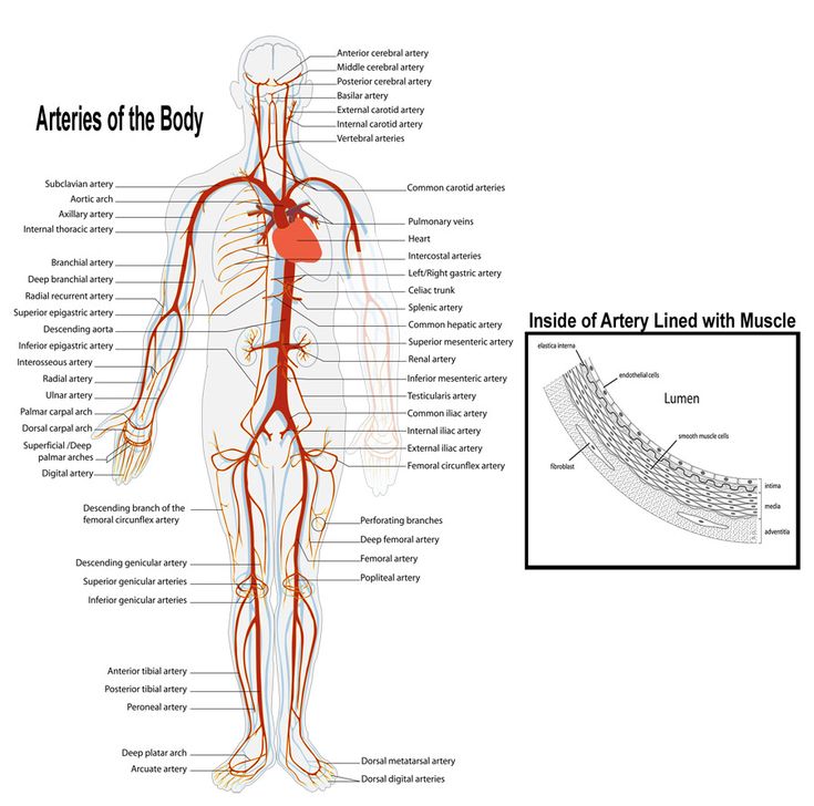
The Arteries of the Body: Structure, Function, and Clinical Relevance

Abstract
The arteries are a critical component of the circulatory system, responsible for transporting oxygenated blood from the heart to the tissues of the body. Arteries are part of a complex network that ensures the delivery of oxygen and nutrients to organs and tissues while also aiding in the removal of waste products. This article provides an expansive overview of the anatomy, function, and clinical relevance of the arterial system.
Introduction
The arterial system forms the first segment of the vascular tree, distributing oxygen-rich blood from the heart to the rest of the body. Arteries are characterized by thick, muscular walls that can withstand the high pressure generated by the heart’s contractions. Understanding the anatomy and function of the arteries is fundamental in medical fields such as cardiology, vascular surgery, and internal medicine.
Structure of Arteries
Arteries are composed of three main layers, each playing a vital role in maintaining the structure and function of the arterial walls:
- Tunica Intima (Inner Layer): This is the innermost layer, consisting of a single layer of endothelial cells that provide a smooth surface for blood flow and regulate vascular tone.
- Tunica Media (Middle Layer): Composed primarily of smooth muscle and elastic fibers, this layer provides strength and elasticity, allowing the arteries to expand and contract with each heartbeat.
- Tunica Externa (Outer Layer): Made up of connective tissue, this layer provides additional structural support and anchors the artery to surrounding tissues.
Types of Arteries
Arteries can be divided into three main types based on their size and function:
- Elastic Arteries: Also known as conducting arteries, these large vessels include the aorta and its major branches. Their elastic walls allow them to stretch and accommodate the surge of blood from the heart during systole (heart contraction) and recoil during diastole (heart relaxation), helping to maintain blood pressure.
- Muscular Arteries: Also called distributing arteries, these vessels include the radial, femoral, and coronary arteries. They contain a higher proportion of smooth muscle in the tunica media and are responsible for distributing blood to specific regions of the body.
- Arterioles: The smallest type of arteries, arterioles lead directly into capillary beds. They play a key role in regulating blood flow and pressure by constricting or dilating in response to various stimuli.
Major Arteries of the Body
The arterial system can be divided into several regions, each supplying different parts of the body.
Arteries of the Head and Neck
- Carotid Arteries:
- Common Carotid Arteries: Arise from the aortic arch (left side) and brachiocephalic trunk (right side), supplying blood to the head and neck.
- Internal Carotid Arteries: Supply oxygenated blood to the brain and eyes.
- External Carotid Arteries: Supply blood to the face, scalp, and neck.
- Vertebral Arteries: Branch from the subclavian arteries and travel through the transverse foramina of the cervical vertebrae to supply blood to the brainstem, cerebellum, and posterior part of the brain.
Arteries of the Upper Limb
- Subclavian Arteries: Supply blood to the upper limbs, neck, and thoracic wall. They give rise to several branches, including the vertebral artery and internal thoracic artery.
- Axillary Arteries: Continuations of the subclavian arteries, supplying blood to the shoulder and upper arm.
- Brachial Arteries: Continuations of the axillary arteries, running along the upper arm and branching into the radial and ulnar arteries at the elbow.
- Radial and Ulnar Arteries: Supply blood to the forearm and hand. The radial artery is commonly used to measure pulse.
Arteries of the Thorax
- Aorta:
- Ascending Aorta: Arises from the left ventricle of the heart, giving rise to the coronary arteries.
- Aortic Arch: Gives rise to the brachiocephalic trunk, left common carotid artery, and left subclavian artery.
- Descending Thoracic Aorta: Continues through the thoracic cavity, giving off branches that supply the chest wall and lungs.
- Intercostal Arteries: Branch from the descending thoracic aorta, supplying the ribs and intercostal muscles.
- Internal Thoracic Arteries: Branch from the subclavian arteries, supplying the anterior chest wall and breasts.
Arteries of the Abdomen
- Abdominal Aorta: Continuation of the descending thoracic aorta after it passes through the diaphragm, supplying blood to the abdominal organs. It gives rise to several key branches:
- Celiac Trunk: Supplies blood to the stomach, liver, spleen, and pancreas.
- Superior Mesenteric Artery: Supplies blood to the small intestine and the first part of the large intestine.
- Inferior Mesenteric Artery: Supplies blood to the distal part of the large intestine.
- Renal Arteries: Supply blood to the kidneys.
- Gonadal Arteries: Supply blood to the testes in males and ovaries in females.
- Common Iliac Arteries: Arise from the bifurcation of the abdominal aorta, supplying the pelvis and lower limbs.
Arteries of the Pelvis and Lower Limb
- Internal Iliac Arteries: Supply blood to the pelvic organs, including the bladder, uterus, and rectum.
- External Iliac Arteries: Continue into the lower limbs, becoming the femoral arteries.
- Femoral Arteries: Supply blood to the thigh, giving rise to the deep femoral artery (which supplies the deeper muscles of the thigh).
- Popliteal Arteries: Continuation of the femoral arteries behind the knee, supplying the knee joint and lower leg.
- Anterior and Posterior Tibial Arteries: Supply blood to the anterior and posterior compartments of the leg, respectively.
- Dorsalis Pedis Artery: A continuation of the anterior tibial artery, supplying blood to the foot and commonly used for assessing the pulse in the foot.
Function of Arteries
The primary function of arteries is to transport oxygenated blood from the heart to the various tissues and organs of the body. This is achieved through the following mechanisms:
- Pressure Regulation: The elastic properties of large arteries, particularly the aorta, allow them to maintain blood pressure and ensure continuous blood flow during both systole and diastole.
- Distribution of Blood: Muscular arteries distribute blood to specific regions of the body, regulating the flow based on the metabolic needs of tissues.
- Vasoconstriction and Vasodilation: Arterioles can constrict or dilate to regulate blood flow into capillary beds, ensuring proper tissue perfusion and blood pressure regulation.
Clinical Relevance
Disorders affecting the arterial system can have significant clinical implications, often leading to life-threatening conditions.
- Atherosclerosis: A condition characterized by the buildup of plaque in the arterial walls, leading to narrowing and reduced blood flow. This can result in coronary artery disease, peripheral artery disease, and stroke.
- Aneurysm: A localized dilation of an artery due to the weakening of its wall. Aneurysms can rupture, causing life-threatening internal bleeding. Common sites include the aorta and cerebral arteries.
- Peripheral Artery Disease (PAD): A condition in which narrowed arteries reduce blood flow to the limbs, often causing pain during walking (claudication) and increasing the risk of infections and ulcers in the feet.
- Hypertension: Chronic high blood pressure can damage arterial walls, leading to complications such as stroke, heart attack, and kidney failure.
- Coronary Artery Disease (CAD): Blockage of the coronary arteries due to atherosclerosis can lead to ischemia of the heart muscle, potentially causing angina or a myocardial infarction (heart attack).
- Carotid Artery Stenosis: Narrowing of the carotid arteries due to atherosclerosis can increase the risk of stroke.
Diagnostic and Therapeutic Approaches
The diagnosis of arterial disorders typically involves imaging techniques, blood pressure monitoring, and functional tests to assess blood flow.
- Doppler Ultrasound: A non-invasive imaging technique used to assess blood flow through arteries and detect blockages or narrowing.
- Angiography: An imaging technique that uses X-rays and contrast material to visualize arteries and identify areas of stenosis or aneurysms.
- Blood Pressure Monitoring: Regular monitoring of blood pressure helps diagnose hypertension, which can contribute to arterial damage.
- Treatment Options:
- Medications: Antihypertensives, statins, and antiplatelet drugs are commonly used to manage arterial disorders.
- Angioplasty and Stenting: A minimally invasive procedure used to open narrowed arteries and restore blood flow.
- Bypass Surgery: In cases of severe arterial blockages, a bypass graft can be used to reroute blood flow around the obstruction.
Conclusion
The arteries of the body are vital for maintaining the flow of oxygenated blood to tissues and organs. Understanding their structure, function, and associated disorders is crucial for the prevention and treatment of cardiovascular diseases. Advances in diagnostic and therapeutic techniques continue to improve the management of arterial conditions, reducing morbidity and mortality associated with arterial diseases.
References
- Standring, S. (2020). Gray’s Anatomy: The Anatomical Basis of Clinical Practice (42nd ed.). Elsevier.
- Moore, K. L., Dalley, A. F., & Agur, A. M. R. (2013). Clinically Oriented Anatomy (7th ed.). Lippincott Williams & Wilkins.
- Netter, F. H. (2014). Atlas of Human Anatomy (6th ed.). Elsevier.
- Williams, P. L., & Warwick, R. (1980). Gray’s Anatomy (36th ed.). Churchill Livingstone.
- Yusuf, S., Reddy, S., Ounpuu, S., & Anand, S. (2001). Global burden of cardiovascular diseases: part I: general considerations, the epidemiologic transition, risk factors, and impact of urbanization. Circulation, 104(22), 2746-2753.
This comprehensive exploration of the arteries of the body highlights their complexity and importance, emphasizing the need for ongoing research and education in cardiovascular health and disease management.
🎓 Want to become a certified instructor?
This lesson is part of our FREE Anatomy course. Create a free account to track your progress and earn your certificate!