The Bones of the Upper Limb: Structure, Function, and Clinical Relevance
Abstract
The upper limb — including the shoulder, arm, forearm, and hand — is one of the most finely tuned systems in orthopedic anatomy. With a total of 30 bones (the clavicle, scapula, humerus, radius, ulna, and hand bones), it supports strength, precision, and functional mobility. This article explores their structure, function, and clinical relevance, emphasizing rehabilitation therapy, fracture management, and musculoskeletal health.
Introduction
The bones of the upper limb provide the essential framework for movement, dexterity, and load-bearing. Understanding their anatomy is vital in medical education, physical therapy, and sports medicine. Accurate anatomical knowledge improves diagnosis and treatment of fractures, dislocations, and degenerative pathologies, and enhances recovery outcomes in rehabilitation and ergonomic therapy.
 Figure 1. Overview of upper limb bones.
Source: Wikimedia Commons, public domain.
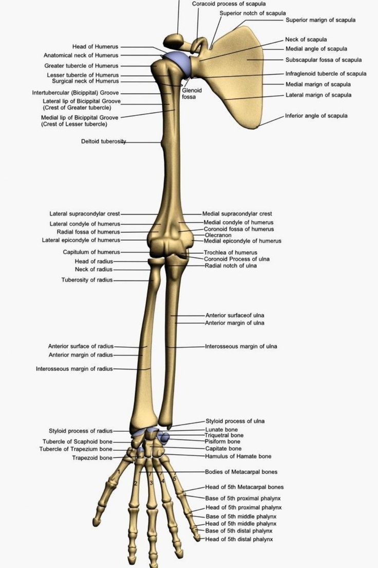
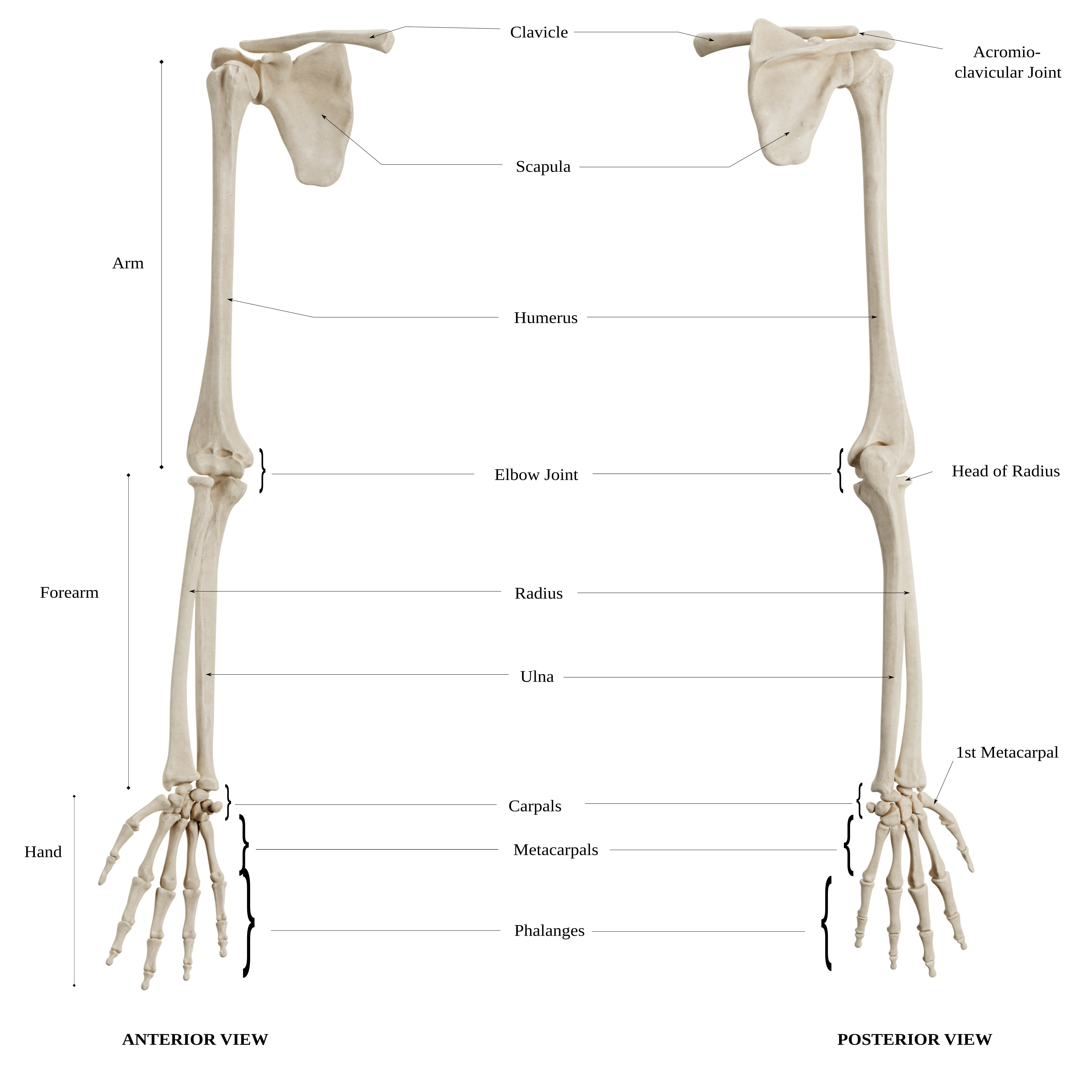 Figure 2. Bones of the upper limb with labeled structures.
Source: Wikimedia Commons, CC BY-SA 4.0.
Anatomical Structure
The upper limb divides into four regions — shoulder girdle, arm, forearm, and hand — each with specific structural and functional roles.
Shoulder Girdle
The shoulder girdle connects the upper limb to the axial skeleton, consisting of:
- Clavicle (Collarbone)
- Scapula (Shoulder Blade)
Clavicle
A curved, S-shaped bone acting as a strut between the sternum and scapula.
- Medial (Sternal) End: Articulates with the manubrium of the sternum.
- Lateral (Acromial) End: Articulates with the acromion of the scapula.
- Functions: Supports the shoulder joint, transmits mechanical forces, protects neurovascular structures, and provides muscle attachment.
 Figure 3. The clavicle (anterior view).
Source: Wikimedia Commons, CC BY-SA 3.0.
Scapula
A flat, triangular bone on the posterior thoracic wall.
- Spine: Divides posterior surface into supraspinous and infraspinous fossae.
- Acromion: Lateral projection forming the top of the shoulder.
- Coracoid Process: Attachment site for muscles and ligaments.
- Glenoid Cavity: Articulates with the humerus forming the glenohumeral joint — a common site of shoulder dislocation.
 Figure 4. Posterior view of the scapula.
Source: Wikimedia Commons, CC BY-SA 3.0.
Arm (Brachium)
The humerus is the only bone in the arm, extending from the shoulder to the elbow.
Humerus
- Head: Articulates with the glenoid cavity.
- Greater/Lesser Tubercles: Attachment sites for rotator cuff muscles.
- Deltoid Tuberosity: Insertion of the deltoid muscle.
- Epicondyles: Anchor points for forearm muscles.
- Capitulum & Trochlea: Form the elbow joint with the radius and ulna.
- Clinical Note: Humeral fractures may affect the radial nerve, requiring orthopedic and physiotherapy intervention.
 Figure 5. Humerus (anterior view).
Source: Wikimedia Commons, CC BY-SA 4.0.
Forearm (Antebrachium)
The forearm contains two bones: the radius (lateral) and ulna (medial), working together for rotation and stability.
Radius
- Head: Articulates with the capitulum and radial notch.
- Radial Tuberosity: Insertion for biceps brachii — important in rehabilitation training.
- Styloid Process: Stabilizes wrist motion.
Ulna
- Olecranon: Forms the elbow’s bony prominence.
- Trochlear Notch: Articulates with the humerus.
- Styloid Process: Attachment for wrist ligaments.
- Clinical Note: Ulnar fractures and nerve entrapment often occur in sports injuries.
 Figure 6. Radius and ulna (posterior view).
Source: Wikimedia Commons, public domain.
Hand
The hand includes the carpus, metacarpus, and phalanges, forming a system of strength and precision.
Carpals (Wrist Bones)
Eight small bones arranged in two rows:
- Proximal – Scaphoid, Lunate, Triquetrum, Pisiform
- Distal – Trapezium, Trapezoid, Capitate, Hamate
Clinical Insight: The scaphoid is the most commonly fractured carpal bone, especially in falls.
Metacarpals (Palm Bones)
Five bones supporting the palm — key for grip strength, fine motor control, and functional therapy.
Phalanges (Finger Bones)
Each finger (except the thumb) has three phalanges: proximal, middle, and distal.
The thumb has two.
Finger fractures and dislocations are common in sports medicine and ergonomic assessment.
 Figure 7. Bones of the hand and wrist.
Source: Wikimedia Commons, CC BY-SA 3.0.
Development and Ossification
Upper limb bones form through endochondral ossification:
- Mesenchymal condensation → cartilage templates
- Primary ossification centers → diaphysis (fetal life)
- Secondary centers → epiphyses (childhood)
Defects may cause growth plate injuries or congenital deformities, studied in pediatric orthopedics.
Function
- Support – Framework for muscles
- Movement – Enables rotation, flexion, extension, abduction
- Protection – Guards nerves and vessels
- Dexterity – Essential for fine motor skills, rehabilitation, and therapy
Clinical Relevance
Common conditions:
- Fractures – clavicle, humerus, radius, or scaphoid
- Dislocations – shoulder and elbow joints
- Arthritis – shoulder, elbow, wrist degeneration
- Carpal Tunnel Syndrome – compression of the median nerve
- Osteoporosis – weakened bone structure
- Tendinopathies – repetitive strain injuries
Treatment often involves imaging, rehabilitation, and surgical care.
Diagnostic and Therapeutic Approaches
- Imaging – X-ray, CT, MRI
- Rehabilitation Therapy – Mobility and strength restoration
- Pharmacologic Management – Pain, inflammation, and bone support
- Surgery – Fixation, arthroplasty, decompression
- Orthotics – Splints and support devices for recovery
Related Clinical Applications
- Critical for orthopedic surgeons, rehabilitation specialists, and physiotherapists
- Central in medical anatomy and biomechanics courses
- Basis for prosthetics, bone density testing, and arthritis innovation
Conclusion
The bones of the upper limb are essential to motion, stability, and strength. Mastery of their anatomy enhances fracture treatment, rehabilitation outcomes, and musculoskeletal health. Modern advances in orthopedic implants, regenerative medicine, and biomechanical design promise faster recovery and improved patient care.
References
- Standring, S. (2020). Gray’s Anatomy: The Anatomical Basis of Clinical Practice (42nd ed.). Elsevier.
- Moore, K. L., Dalley, A. F., & Agur, A. M. R. (2013). Clinically Oriented Anatomy (7th ed.). Lippincott Williams & Wilkins.
- Sadler, T. W. (2018). Langman’s Medical Embryology (14th ed.). Wolters Kluwer.
- Netter, F. H. (2014). Atlas of Human Anatomy (6th ed.). Elsevier.
- Rockwood, C. A., Green, D. P., & Bucholz, R. W. (2010). Rockwood and Green’s Fractures in Adults (7th ed.). Lippincott Williams & Wilkins.
🎓 Want to become a certified instructor?
This lesson is part of our FREE Anatomy course. Create a free account to track your progress and earn your certificate!