The Heart: Structure, Function, and Clinical Relevance

Abstract
The heart is the central organ of the circulatory system, responsible for pumping blood throughout the body to supply oxygen and nutrients while removing waste products. It functions as a double pump, delivering deoxygenated blood to the lungs and oxygenated blood to the rest of the body. This article provides an in-depth overview of the anatomy, function, and clinical significance of the heart, highlighting its role in maintaining homeostasis and supporting life.
Introduction
The heart is a muscular organ located in the thoracic cavity, specifically within the mediastinum, between the lungs. As the core of the cardiovascular system, the heart continuously pumps blood through two major circulatory pathways: the pulmonary circulation (to the lungs) and the systemic circulation (to the body). Its coordinated contractions ensure that oxygen-rich blood reaches all tissues while carbon dioxide and metabolic waste products are transported away for excretion.
Anatomy of the Heart
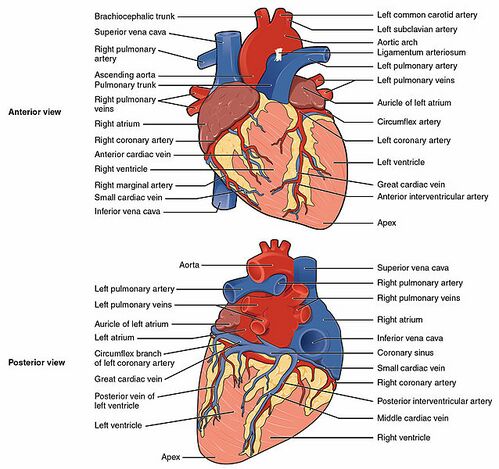
The heart is structurally complex, consisting of chambers, valves, vessels, and a conductive system that regulates its rhythmic contractions. It is surrounded by a protective sac called the pericardium, which helps anchor the heart and prevents friction during movement.
1. Chambers of the Heart
The heart has four chambers: two atria (upper chambers) and two ventricles (lower chambers), separated by septa that prevent the mixing of oxygenated and deoxygenated blood.
- Right Atrium: Receives deoxygenated blood from the body via the superior and inferior vena cava. Blood passes through the tricuspid valve into the right ventricle.
- Right Ventricle: Pumps deoxygenated blood into the pulmonary arteries, which carry blood to the lungs for oxygenation.
- Left Atrium: Receives oxygenated blood from the lungs via the pulmonary veins. Blood flows through the mitral valve into the left ventricle.
- Left Ventricle: The most muscular chamber, it pumps oxygenated blood into the aorta for distribution throughout the body. It generates the highest pressure of any chamber due to its role in systemic circulation.
2. Heart Valves
Heart valves ensure unidirectional blood flow through the heart’s chambers and prevent backflow during contraction.
- Atrioventricular Valves:
- Tricuspid Valve: Located between the right atrium and right ventricle, it prevents backflow of blood into the atrium when the ventricle contracts.
- Mitral (Bicuspid) Valve: Located between the left atrium and left ventricle, it ensures that blood flows only into the ventricle and not backward into the atrium.
- Semilunar Valves:
- Pulmonary Valve: Between the right ventricle and the pulmonary artery, it prevents backflow of blood into the ventricle after it has been pumped to the lungs.
- Aortic Valve: Between the left ventricle and the aorta, it prevents blood from flowing back into the ventricle after being pumped into the systemic circulation.
3. Layers of the Heart Wall
The heart wall consists of three distinct layers, each with a unique function:
- Endocardium: The innermost layer, composed of a thin layer of endothelial cells, which lines the chambers of the heart and is continuous with the blood vessels.
- Myocardium: The thick, muscular middle layer responsible for the contractile force that pumps blood. It contains cardiac muscle fibers, which are highly specialized for endurance and rhythmic contractions.
- Epicardium: The outer layer, also known as the visceral pericardium, which provides structural support and lubrication to reduce friction as the heart beats.
4. Coronary Circulation
The heart is supplied with oxygen and nutrients by the coronary arteries, which branch off the aorta. There are two main coronary arteries:
- Left Coronary Artery: Branches into the left anterior descending (LAD) artery and the circumflex artery, supplying the left side of the heart.
- Right Coronary Artery: Supplies the right side of the heart and branches into the posterior descending artery.
Oxygen-depleted blood from the heart muscle is returned via coronary veins into the coronary sinus, which empties into the right atrium.
Function of the Heart
The primary function of the heart is to pump blood through the pulmonary and systemic circulations, maintaining the flow of oxygenated and deoxygenated blood throughout the body. This process is cyclical and consists of two main phases: systole and diastole.
1. Cardiac Cycle
The cardiac cycle refers to the sequence of events that occur during one heartbeat and is divided into:
- Systole: The contraction phase of the heart, during which the ventricles contract, forcing blood into the pulmonary arteries (right ventricle) and aorta (left ventricle).
- Diastole: The relaxation phase of the heart, during which the ventricles fill with blood from the atria in preparation for the next contraction.
The heart’s ability to pump blood efficiently depends on its electrical conduction system, which coordinates the timing of atrial and ventricular contractions.
2. Electrical Conduction System
The heart has an intrinsic electrical system that controls its rhythmic contractions. The key components include:
- Sinoatrial (SA) Node: Often called the heart’s "pacemaker," it generates electrical impulses that initiate each heartbeat. The SA node is located in the right atrium and causes the atria to contract, pushing blood into the ventricles.
- Atrioventricular (AV) Node: Located between the atria and ventricles, the AV node delays the electrical impulse to ensure that the ventricles fill with blood before they contract.
- Bundle of His: A group of fibers that transmits the electrical signal from the AV node to the ventricles.
- Purkinje Fibers: Specialized fibers that distribute the electrical impulse to the ventricular muscle, causing the ventricles to contract and pump blood.
The normal heart rate is regulated by this conduction system, typically generating 60 to 100 beats per minute at rest.
Regulation of Heart Function
The heart’s activity is influenced by both intrinsic factors and external inputs, including neural and hormonal signals.
- Autonomic Nervous System (ANS): The ANS regulates heart rate and force of contraction.
- Sympathetic Stimulation: Increases heart rate and contractility, preparing the body for "fight or flight" responses.
- Parasympathetic Stimulation: Decreases heart rate and promotes "rest and digest" functions through the vagus nerve.
- Hormones: Epinephrine and norepinephrine, released during stress or exercise, increase heart rate and contractility. Hormones such as thyroid hormones also affect heart function.
Clinical Relevance
The heart is subject to various disorders and diseases that can significantly impact overall health and quality of life. Common heart-related conditions include:
- Coronary Artery Disease (CAD): A condition caused by the buildup of atherosclerotic plaques in the coronary arteries, reducing blood flow to the heart muscle. It can lead to angina (chest pain) or a myocardial infarction (heart attack).
- Heart Failure: A condition in which the heart cannot pump blood efficiently, leading to fluid buildup in the lungs and extremities. It can result from conditions such as hypertension, heart attacks, or valvular disease.
- Arrhythmias: Abnormal heart rhythms caused by disruptions in the electrical conduction system. Arrhythmias can range from benign (e.g., premature atrial contractions) to life-threatening (e.g., ventricular fibrillation).
- Valvular Heart Disease: A condition in which one or more of the heart valves fail to function properly. Common problems include stenosis (narrowing of the valve) and regurgitation (leakage of the valve).
- Hypertrophic Cardiomyopathy: A genetic condition characterized by the thickening of the heart muscle, often leading to obstruction of blood flow and an increased risk of sudden cardiac death.
- Pericarditis: Inflammation of the pericardium, often causing chest pain and fluid accumulation around the heart.
Diagnostic and Therapeutic Approaches
Several diagnostic tools and treatment options are available to assess and manage heart conditions:
- Electrocardiogram (ECG): A test that records the electrical activity of the heart, commonly used to diagnose arrhythmias, heart attacks, and other heart conditions.
- Echocardiography: An ultrasound of the heart that provides detailed images of the heart’s structure, including the valves and chambers, as well as blood flow.
- Cardiac Catheterization: A procedure in which a catheter is inserted into a coronary artery to diagnose and treat blockages. It is often combined with angioplasty and stenting to open narrowed arteries.
- Medications: Common medications used in heart disease management include:
- Beta-blockers: Reduce heart rate and blood pressure.
- ACE inhibitors: Lower blood pressure and decrease strain on the heart.
- Anticoagulants: Prevent blood clot formation.
- Statins: Lower cholesterol levels.
- Coronary Artery Bypass Grafting (CABG): A surgical procedure in which blood flow is rerouted around blocked coronary arteries using grafts from other blood vessels.
- Implantable Devices: Pacemakers and implantable cardioverter-defibrillators (ICDs) are used to regulate abnormal heart rhythms and prevent sudden cardiac death.
Conclusion
The heart is a vital organ that ensures the continuous circulation of blood throughout the body, delivering oxygen and nutrients to tissues and removing waste products. Understanding its anatomy, function, and associated clinical conditions is crucial for diagnosing and managing cardiovascular diseases. Advances in diagnostic techniques, medications, and surgical interventions have greatly improved the prognosis for individuals with heart disease, enhancing both survival and quality of life.
References
- Standring, S. (2020). Gray’s Anatomy: The Anatomical Basis of Clinical Practice (42nd ed.). Elsevier.
- Lilly, L. S. (2015). Pathophysiology of Heart Disease (6th ed.). Wolters Kluwer.
- Braunwald, E. (2011). Heart Disease: A Textbook of Cardiovascular Medicine (9th ed.). Elsevier.
- Fuster, V., & Harrington, R. A. (2017). Hurst’s the Heart (14th ed.). McGraw-Hill.
- Moore, K. L., Dalley, A. F., & Agur, A. M. R. (2013). Clinically Oriented Anatomy (7th ed.). Lippincott Williams & Wilkins.
This detailed exploration of the heart underscores its essential role in sustaining life and highlights the importance of ongoing research and medical advancements in cardiology.
🎓 Want to become a certified instructor?
This lesson is part of our FREE Anatomy course. Create a free account to track your progress and earn your certificate!