The Muscles of the Lower Extremity: Structure, Function, and Clinical Relevance
Abstract
The muscles of the lower extremity, comprising the hip, thigh, leg, and foot, are essential for locomotion, balance, and support of the body’s weight. These muscles are categorized based on their anatomical location and function, facilitating a wide range of movements necessary for walking, running, jumping, and other activities. This article provides an in-depth examination of the anatomy, development, functions, and clinical significance of the muscles of the lower extremity.
Introduction
The lower extremity muscles are fundamental to human movement and mobility. They enable activities such as standing, walking, running, and jumping, while also playing a critical role in maintaining balance and posture. Understanding the anatomy and function of these muscles is crucial in fields such as orthopedics, physical therapy, sports medicine, and rehabilitation.
Anatomical Structure
The muscles of the lower extremity can be categorized into four main groups based on their location and function:
- Muscles of the Hip
- Muscles of the Thigh
- Muscles of the Leg
- Muscles of the Foot
Muscles of the Hip
The muscles of the hip are responsible for the movement and stabilization of the hip joint. They can be divided into gluteal muscles, lateral rotator muscles, and iliopsoas muscles.
Gluteal Muscles
These muscles are located on the posterior aspect of the hip and are crucial for the movement and stability of the hip joint.
- Gluteus Maximus: The largest and most superficial of the gluteal muscles, it extends and externally rotates the hip; also involved in maintaining an upright posture.
- Gluteus Medius: Located deep to the gluteus maximus, it abducts and medially rotates the hip; essential for stabilizing the pelvis during walking.
- Gluteus Minimus: The smallest of the gluteal muscles, located deep to the gluteus medius, it also abducts and medially rotates the hip.
- Tensor Fasciae Latae: Assists in abduction and medial rotation of the hip; also plays a role in stabilizing the knee through the iliotibial tract.
Lateral Rotator Muscles
These muscles are responsible for the external rotation of the hip and include:
- Piriformis: Externally rotates and abducts the hip.
- Obturator Internus: Externally rotates the hip.
- Obturator Externus: Externally rotates the hip.
- Superior and Inferior Gemellus: Assist in external rotation of the hip.
- Quadratus Femoris: Externally rotates and stabilizes the hip.
Iliopsoas Muscles
The iliopsoas is the primary flexor of the hip and consists of two muscles:
- Psoas Major: Flexes the hip and stabilizes the lumbar spine.
- Iliacus: Works in conjunction with the psoas major to flex the hip.
Muscles of the Thigh
The thigh muscles are divided into three compartments: anterior, medial, and posterior, each containing muscles that perform specific functions.
Anterior Compartment
These muscles are primarily involved in the extension of the knee and flexion of the hip.
- Quadriceps Femoris: The largest muscle group in the anterior compartment, consisting of four muscles:
- Rectus Femoris: Extends the knee and flexes the hip.
- Vastus Lateralis: Extends the knee.
- Vastus Medialis: Extends the knee.
- Vastus Intermedius: Extends the knee.
- Sartorius: The longest muscle in the human body, it flexes, abducts, and laterally rotates the hip, and flexes the knee.
Medial Compartment
These muscles are primarily involved in the adduction of the thigh.
- Adductor Longus: Adducts and medially rotates the thigh.
- Adductor Brevis: Adducts and medially rotates the thigh.
- Adductor Magnus: The largest adductor muscle, it adducts, flexes, and extends the thigh.
- Gracilis: A long, slender muscle that adducts the thigh and flexes the knee.
- Pectineus: Adducts and flexes the thigh.
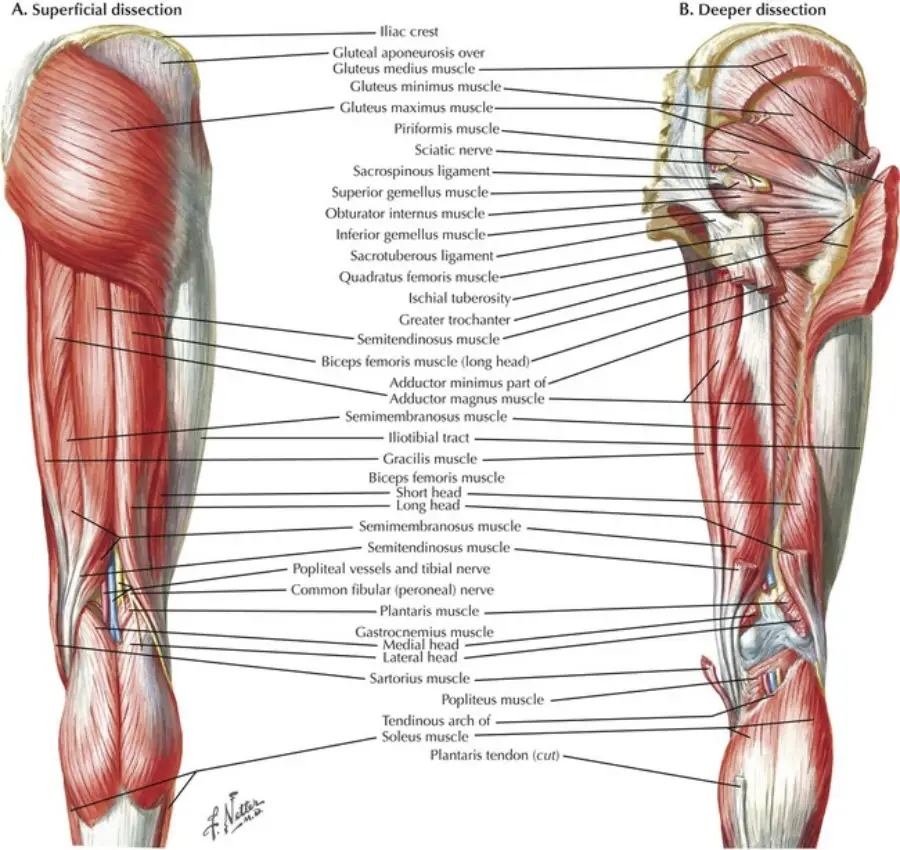
Posterior Compartment
These muscles, also known as the hamstrings, are primarily involved in the flexion of the knee and extension of the hip.
- Biceps Femoris: Flexes the knee and extends the hip.
- Semitendinosus: Flexes the knee, extends the hip, and medially rotates the leg.
- Semimembranosus: Flexes the knee, extends the hip, and medially rotates the leg.
Muscles of the Leg
The leg muscles are divided into three compartments: anterior, lateral, and posterior, each containing muscles that perform specific functions.
Anterior Compartment
These muscles are primarily involved in dorsiflexion of the foot and extension of the toes.
- Tibialis Anterior: Dorsiflexes and inverts the foot.
- Extensor Digitorum Longus: Extends the toes and dorsiflexes the foot.
- Extensor Hallucis Longus: Extends the big toe and dorsiflexes the foot.
- Fibularis (Peroneus) Tertius: Dorsiflexes and everts the foot.
Lateral Compartment
These muscles are primarily involved in eversion and plantarflexion of the foot.
- Fibularis (Peroneus) Longus: Everts and plantarflexes the foot; also supports the arch of the foot.
- Fibularis (Peroneus) Brevis: Everts and plantarflexes the foot.
Posterior Compartment
The posterior compartment is further divided into superficial and deep layers, with muscles involved in plantarflexion of the foot and flexion of the toes.
- Superficial Layer:
- Gastrocnemius: A two-headed muscle that plantarflexes the foot and flexes the knee.
- Soleus: Located deep to the gastrocnemius, it plantarflexes the foot.
- Plantaris: A small muscle that assists in plantarflexion of the foot.
- Deep Layer:
- Popliteus: Unlocks the knee by rotating the femur on the tibia.
- Flexor Hallucis Longus: Flexes the big toe and assists in plantarflexion of the foot.
- Flexor Digitorum Longus: Flexes the toes and assists in plantarflexion of the foot.
- Tibialis Posterior: Inverts and plantarflexes the foot; also supports the arch of the foot.
Muscles of the Foot
The muscles of the foot are responsible for the fine motor movements of the toes and help maintain the arches of the foot. They are divided into intrinsic and extrinsic muscles.
Intrinsic Muscles
These muscles are located entirely within the foot and are involved in movements of the toes and maintenance of foot arches.
- Dorsal Group:
- Extensor Digitorum Brevis: Extends the toes.
- Extensor Hallucis Brevis: Extends the big toe.
- Plantar Group:
- Abductor Hallucis: Abducts the big toe.
- Flexor Digitorum Brevis: Flexes the middle phalanges of the toes.
- Abductor Digiti Minimi: Abducts the little toe.
- Quadratus Plantae: Assists in flexing the toes.
- Lumbricals: Flex the metatarsophalangeal joints and extend the interphalangeal joints.
- Flexor Hallucis Brevis: Flexes the big toe.
- Adductor Hallucis: Adducts the big toe.
- Flexor Digiti Minimi Brevis: Flexes the little toe.
- Plantar Interossei: Adduct the toes.
- Dorsal Interossei: Abduct the toes.
Extrinsic Muscles
These muscles originate in the leg and insert on the foot, controlling gross movements of the toes and foot.
- Flexor Digitorum Longus: Flexes the toes and assists in plantarflexion of the foot.
- Flexor Hallucis Longus: Flexes the big toe and assists in plantarflexion of the foot.
- Extensor Digitorum Longus: Extends the toes and dorsiflexes the foot.
- Extensor Hallucis Longus: Extends the big toe and dorsiflexes the foot.
Development
The muscles of the lower extremity develop from the paraxial mesoderm, specifically from the somites. The somites differentiate into myotomes, which further divide into dorsal (epaxial) and ventral (hypaxial) portions, giving rise to the muscles of the lower extremity.
- Epaxial Myotomes: Contribute to the development of the back muscles.
Hypaxial Myotomes: Develop into the limb muscles, including those of the lower extremity.
Function
The muscles of the lower extremity serve several essential functions:
- Locomotion: Facilitate walking, running, jumping, and other forms of movement.
- Support and Posture: Maintain the body’s upright posture and balance.
- Force Generation: Enable powerful movements such as lifting, pushing, and climbing.
- Stabilization: Provide stability to the joints of the hip, knee, and ankle during movement.
- Shock Absorption: Help absorb the impact during activities like walking and running.
Clinical Relevance
Disorders and injuries affecting the muscles of the lower extremity can lead to significant clinical problems:
- Hamstring Strain: A common injury in athletes, leading to pain and limited movement in the posterior thigh.
- Quadriceps Tendinitis: Inflammation of the quadriceps tendon, causing pain in the anterior thigh and knee.
- Achilles Tendinitis: Inflammation of the Achilles tendon, leading to pain and stiffness in the heel.
- Plantar Fasciitis: Inflammation of the plantar fascia, causing pain in the heel and arch of the foot.
- Iliotibial Band Syndrome: A common overuse injury affecting the lateral knee, often seen in runners and cyclists.
- Piriformis Syndrome: Compression of the sciatic nerve by the piriformis muscle, leading to pain and numbness in the buttocks and lower extremity.
- Shin Splints (Medial Tibial Stress Syndrome): Pain along the inner edge of the tibia, commonly caused by overuse or improper footwear.
- Foot Drop: Weakness or paralysis of the muscles involved in lifting the front part of the foot, leading to difficulty walking.
Diagnostic and Therapeutic Approaches
Diagnosis of lower extremity muscle disorders typically involves clinical examination, imaging techniques (such as X-rays, MRI, and ultrasound), and electromyography (EMG) to assess muscle function. Treatment options vary depending on the condition and may include:
- Physical Therapy: Exercises to strengthen and stretch the lower extremity muscles, improve flexibility, and alleviate pain.
- Medications: Pain relievers, anti-inflammatory drugs, and corticosteroid injections.
- Manual Therapy: Techniques such as massage, joint mobilization, and trigger point therapy.
- Surgical Interventions: In severe cases, such as Achilles tendon rupture or severe plantar fasciitis, surgery may be necessary.
- Orthotic Devices: Custom-made shoe inserts to support the arches of the foot and correct alignment issues.
- Rest and Ice Therapy: Common initial treatments for acute muscle injuries to reduce inflammation and pain.
Conclusion
The muscles of the lower extremity are vital for movement, support, and balance, enabling humans to perform a wide range of activities, from basic walking to complex athletic maneuvers. Understanding their anatomy, development, and potential disorders is crucial for effective medical care. Advances in diagnostic techniques and treatment options continue to improve the management of lower extremity muscle disorders, enhancing patient outcomes and quality of life.
References
- Standring, S. (2020). Gray’s Anatomy: The Anatomical Basis of Clinical Practice (42nd ed.). Elsevier.
- Moore, K. L., Dalley, A. F., & Agur, A. M. R. (2013). Clinically Oriented Anatomy (7th ed.). Lippincott Williams & Wilkins.
- Netter, F. H. (2014). Atlas of Human Anatomy (6th ed.). Elsevier.
- Williams, P. L., & Warwick, R. (1980). Gray’s Anatomy (36th ed.). Churchill Livingstone.
- Bogduk, N. (2005). Clinical Anatomy of the Lumbar Spine and Sacrum (4th ed.). Churchill Livingstone.
- Kendall, F. P., McCreary, E. K., & Provance, P. G. (2005). Muscles: Testing and Function, with Posture and Pain (5th ed.). Lippincott Williams & Wilkins.
This comprehensive exploration of the muscles of the lower extremity highlights their complexity and importance, emphasizing the need for ongoing research and education in musculoskeletal health and disease management.
🎓 Want to become a certified instructor?
This lesson is part of our FREE Anatomy course. Create a free account to track your progress and earn your certificate!