The Neck Muscles: Structure, Function, and Clinical Relevance
Abstract
The neck muscles are a complex and vital group of muscles that play crucial roles in head and neck movement, support, and stabilization. They are also essential for functions such as respiration, swallowing, and maintaining posture. This article provides an in-depth examination of the anatomy, development, functions, and clinical significance of the neck muscles.
Introduction
The neck muscles are involved in a wide range of movements and activities, making them essential for both functional and aesthetic purposes. They can be categorized based on their location and function, including superficial muscles, deep muscles, and muscles associated with the cervical vertebrae, hyoid bone, and thoracic inlet. Understanding the anatomy and function of these muscles is crucial in fields such as neurology, orthopedics, physical therapy, and surgery.
Anatomical Structure
The neck muscles can be divided into several groups based on their location and function:
- Superficial Neck Muscles
- Suprahyoid and Infrahyoid Muscles
- Deep Neck Muscles
- Scalene Muscles
- Prevertebral Muscles
- Muscles Associated with the Cervical Spine
Superficial Neck Muscles
These muscles are located just beneath the skin and are primarily involved in movement and support of the head and neck.
- Platysma: A thin, broad muscle that extends from the lower jaw (mandible) to the upper chest. It is involved in depressing the mandible and tensing the skin of the neck.
- Sternocleidomastoid (SCM): A prominent, paired muscle that runs from the sternum and clavicle to the mastoid process of the temporal bone. The SCM is responsible for flexing and rotating the head, as well as contributing to the elevation of the thoracic cage during respiration.
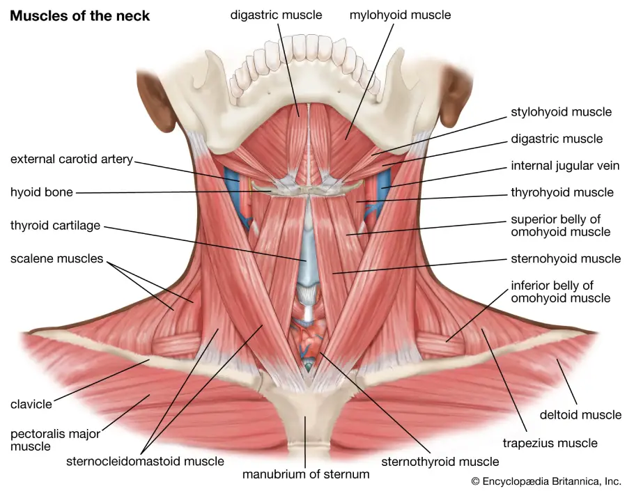
Suprahyoid and Infrahyoid Muscles
These muscles are associated with the hyoid bone and are involved in swallowing and movements of the larynx.
Suprahyoid Muscles
- Mylohyoid: Forms the floor of the mouth and elevates the hyoid bone during swallowing.
- Geniohyoid: Assists in pulling the hyoid bone anteriorly and upward.
- Stylohyoid: Elevates and retracts the hyoid bone.
- Digastric: Composed of two bellies (anterior and posterior), it elevates the hyoid bone and depresses the mandible.
Infrahyoid Muscles
- Sternohyoid: Depresses the hyoid bone after it has been elevated during swallowing.
- Omohyoid: A two-bellied muscle that depresses the hyoid bone.
- Sternothyroid: Depresses the thyroid cartilage.
- Thyrohyoid: Depresses the hyoid bone and elevates the thyroid cartilage.
Deep Neck Muscles
These muscles are located deep within the neck and play a crucial role in stabilizing the cervical spine and facilitating head and neck movements.
- Longus Colli: Runs along the anterior aspect of the cervical vertebrae, flexing and rotating the neck.
- Longus Capitis: Extends from the cervical vertebrae to the occipital bone, flexing the head.
- Rectus Capitis Anterior: A small muscle that assists in flexing the head at the atlanto-occipital joint.
- Rectus Capitis Lateralis: Helps in lateral flexion of the head.
Scalene Muscles
The scalene muscles are a group of three paired muscles (anterior, middle, and posterior) located on the lateral aspect of the neck. They are involved in flexing the neck laterally and elevating the first and second ribs during forced inspiration.
- Anterior Scalene: Originates from the transverse processes of the cervical vertebrae and inserts onto the first rib.
- Middle Scalene: Lies posterior to the anterior scalene, also attaching to the first rib.
- Posterior Scalene: The smallest of the scalene muscles, it attaches to the second rib.
Prevertebral Muscles
These muscles lie directly in front of the vertebral column and are involved in flexing the neck and head.
- Longus Colli: As mentioned earlier, it flexes and slightly rotates the neck.
- Longus Capitis: Flexes the head and neck.
- Rectus Capitis Anterior and Lateralis: Assist in flexing and stabilizing the head.
Muscles Associated with the Cervical Spine
These muscles are associated with the movement and stabilization of the cervical spine.
- Splenius Capitis: Extends and rotates the head.
- Splenius Cervicis: Extends and rotates the cervical spine.
- Semispinalis Capitis: Extends and rotates the head.
- Semispinalis Cervicis: Extends and rotates the cervical spine.
Development
The muscles of the neck develop from the mesodermal layer of the embryo, specifically from the myotomes of the somites, which are segments of the mesoderm located along the developing spinal cord. The neck muscles arise from different groups of myotomes, reflecting their varied functions and innervation.
- Branchial Arch Muscles: The muscles of facial expression and some neck muscles develop from the branchial arches.
- Cervical Myotomes: Give rise to the deeper muscles of the neck, including the scalene and prevertebral muscles.
Function
The neck muscles serve several essential functions:
- Movement: Facilitate flexion, extension, rotation, and lateral bending of the head and neck.
- Support and Stabilization: Maintain the position of the head and neck, particularly during movement and postural changes.
- Respiration: Assist in the elevation of the ribs during forced breathing.
- Swallowing and Speech: Elevate and depress the hyoid bone and larynx, contributing to swallowing and vocalization.
- Protection: Protect vital structures such as the cervical spine, blood vessels, and nerves.
Clinical Relevance
Disorders and injuries affecting the neck muscles can lead to significant clinical problems:
- Cervical Strain: Often results from overuse or acute injury, leading to pain and stiffness in the neck.
- Torticollis (Wry Neck): A condition characterized by an abnormal, asymmetrical head or neck position, often due to muscle spasms.
- Whiplash Injury: A common result of car accidents, involving rapid flexion and extension of the neck, leading to muscle strain and potential ligament damage.
- Thoracic Outlet Syndrome: Compression of neurovascular structures between the scalene muscles and first rib, leading to pain, numbness, and weakness in the upper limb.
- Myofascial Pain Syndrome: Chronic pain in the neck muscles due to trigger points, which are hyperirritable spots in the muscle.
- Degenerative Disc Disease: Age-related changes in the cervical spine can lead to muscle spasms and pain.
Diagnostic and Therapeutic Approaches
Diagnosis of neck muscle disorders often involves clinical examination, imaging techniques such as X-rays, MRI, and CT scans, and electromyography (EMG) to assess muscle function. Treatment varies depending on the condition and may include:
- Physical Therapy: Exercises and stretches to strengthen muscles, improve flexibility, and reduce pain.
- Medications: Pain relievers, muscle relaxants, and anti-inflammatory drugs.
- Manual Therapy: Techniques such as massage, chiropractic adjustments, and trigger point therapy.
- Surgical Interventions: In severe cases, such as with cervical spine instability or persistent thoracic outlet syndrome, surgery may be necessary.
- Posture Correction: Ergonomic adjustments and posture training to prevent and alleviate neck strain.
Conclusion
The muscles of the neck are crucial for a wide range of functions, from supporting and moving the head to aiding in respiration and swallowing. Understanding their anatomy, development, and potential disorders is essential for effective medical care. Advances in diagnostic techniques and treatment options continue to improve the management of neck muscle disorders, enhancing patient outcomes and quality of life.
References
- Standring, S. (2020). Gray’s Anatomy: The Anatomical Basis of Clinical Practice (42nd ed.). Elsevier.
- Moore, K. L., Dalley, A. F., & Agur, A. M. R. (2013). Clinically Oriented Anatomy (7th ed.). Lippincott Williams & Wilkins.
- Netter, F. H. (2014). Atlas of Human Anatomy (6th ed.). Elsevier.
- Williams, P. L., & Warwick, R. (1980). Gray’s Anatomy (36th ed.). Churchill Livingstone.
- Agur, A. M. R., & Dalley, A. F. (2017). Grant’s Atlas of Anatomy (14th ed.). Lippincott Williams & Wilkins.
- Bogduk, N. (2005). Clinical Anatomy of the Lumbar Spine and Sacrum (4th ed.). Churchill Livingstone.
This comprehensive exploration of the neck muscles emphasizes their complexity and importance, highlighting the need for ongoing research and education in neuromuscular health and disease management.
🎓 Want to become a certified instructor?
This lesson is part of our FREE Anatomy course. Create a free account to track your progress and earn your certificate!