The Respiratory System: Structure, Function, and Clinical Relevance
Abstract
The respiratory system is essential for the exchange of oxygen and carbon dioxide between the body and the external environment. It includes the airways, lungs, and muscles of respiration, which work together to facilitate breathing. This article explores the anatomy, function, and clinical significance of the respiratory system, highlighting its critical role in maintaining homeostasis and supporting life.
Introduction
The respiratory system is responsible for delivering oxygen to the body and removing carbon dioxide, a metabolic waste product. This process, known as gas exchange, is vital for cellular respiration, which provides energy for cellular functions. The respiratory system is divided into the upper and lower respiratory tracts and is supported by respiratory muscles. Understanding the structure and function of this system is crucial for diagnosing and treating respiratory disorders.
Structure of the Respiratory System
The respiratory system can be divided into two main sections:
- Upper Respiratory Tract
- Lower Respiratory Tract
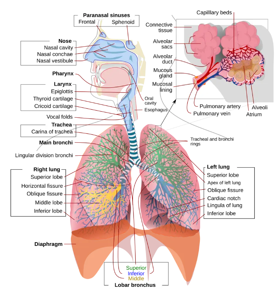
Upper Respiratory Tract
The upper respiratory tract is responsible for filtering, warming, and humidifying air before it reaches the lower respiratory tract. It includes the following structures:
- Nose and Nasal Cavity: The primary entry point for inhaled air. The nasal cavity is lined with mucous membranes and cilia that filter out dust and pathogens.
- Pharynx (Throat): A muscular tube that connects the nasal cavity to the larynx. It serves as a passageway for both air and food.
- Larynx (Voice Box): Located at the top of the trachea, the larynx houses the vocal cords and plays a key role in voice production. It also acts as a gateway that prevents food and liquids from entering the lower respiratory tract during swallowing.
Lower Respiratory Tract
The lower respiratory tract is involved in gas exchange and includes the following structures:
- Trachea (Windpipe): A flexible tube that connects the larynx to the bronchi. It is reinforced with C-shaped cartilaginous rings to prevent collapse during breathing.
- Bronchi: The trachea splits into two primary bronchi, one for each lung. The bronchi further divide into smaller bronchi and bronchioles, creating a branching network within the lungs.
- Bronchioles: Smaller airways that lead to the alveoli. Bronchioles are surrounded by smooth muscle that can constrict or dilate to regulate airflow.
- Alveoli: Tiny air sacs at the end of the bronchioles where gas exchange occurs. The alveoli are surrounded by capillaries, allowing oxygen to diffuse into the blood and carbon dioxide to diffuse out.
Lungs
- Location: The lungs are located on either side of the heart within the thoracic cavity. Each lung is divided into lobes: the right lung has three lobes (superior, middle, and inferior), while the left lung has two lobes (superior and inferior) to accommodate the heart.
- Pleura: The lungs are covered by a double-layered membrane called the pleura. The visceral pleura covers the lungs, and the parietal pleura lines the chest wall. Between the two layers is the pleural cavity, filled with pleural fluid that reduces friction during breathing.
Respiratory Muscles
The primary muscles involved in breathing are the diaphragm and the intercostal muscles:
- Diaphragm: A dome-shaped muscle located below the lungs. When the diaphragm contracts, it flattens, increasing the volume of the thoracic cavity and causing air to be drawn into the lungs.
- Intercostal Muscles: These muscles are located between the ribs. The external intercostals raise the rib cage during inhalation, while the internal intercostals assist in forced exhalation.
Function of the Respiratory System
The primary function of the respiratory system is gas exchange, ensuring that oxygen is delivered to the body while carbon dioxide is expelled. This process is achieved through two key mechanisms:
- Ventilation: The physical process of moving air into and out of the lungs, commonly known as breathing. It is divided into:
- Inhalation (Inspiration): During inhalation, the diaphragm contracts and moves downward, increasing the volume of the thoracic cavity. This causes a drop in pressure inside the lungs, allowing air to flow in.
- Exhalation (Expiration): During exhalation, the diaphragm relaxes and moves upward, decreasing the volume of the thoracic cavity and forcing air out of the lungs. Exhalation is typically passive but can be active during activities such as speaking or exercising.
- Gas Exchange: This occurs at the alveolar level:
- Oxygen Transport: Oxygen from inhaled air diffuses across the alveolar walls into the capillaries, where it binds to hemoglobin in red blood cells and is transported to tissues.
- Carbon Dioxide Removal: Carbon dioxide, a waste product of metabolism, diffuses from the capillaries into the alveoli and is expelled from the body during exhalation.
Other functions of the respiratory system include:
- Acid-Base Balance: The respiratory system helps regulate blood pH by controlling the levels of carbon dioxide, which can affect the concentration of hydrogen ions in the blood.
- Filtration and Immune Defense: The respiratory tract is lined with cilia and mucus that trap and remove dust, pathogens, and other particles. Immune cells in the lungs also help protect against infection.
- Speech Production: The larynx, vocal cords, and air moving through the respiratory system enable vocalization and speech.
Control of Breathing
Breathing is controlled by the respiratory centers located in the brainstem, particularly the medulla oblongata and the pons. These centers regulate the rate and depth of breathing in response to changes in carbon dioxide, oxygen, and blood pH levels detected by chemoreceptors in the aorta, carotid arteries, and brain.
- Medulla Oblongata: Regulates the basic rhythm of breathing by sending signals to the diaphragm and intercostal muscles to contract.
- Pons: Helps fine-tune breathing patterns and prevent over-inflation of the lungs.
Clinical Relevance
Respiratory disorders can significantly affect the ability to breathe and maintain adequate gas exchange. Common conditions include:
- Asthma: A chronic condition characterized by inflammation and narrowing of the airways, leading to wheezing, shortness of breath, and chest tightness. Asthma attacks are often triggered by allergens, exercise, or stress. Treatment includes bronchodilators and anti-inflammatory medications.
- Chronic Obstructive Pulmonary Disease (COPD): A progressive disease that includes emphysema and chronic bronchitis. COPD is usually caused by smoking and leads to obstructed airflow, making it difficult to breathe. Symptoms include shortness of breath, chronic cough, and frequent respiratory infections.
- Pneumonia: An infection that inflames the alveoli in one or both lungs, filling them with fluid or pus. It can be caused by bacteria, viruses, or fungi. Symptoms include fever, cough, and difficulty breathing. Pneumonia is diagnosed through chest X-rays and treated with antibiotics or antivirals.
- Lung Cancer: A malignant tumor in the lungs, often associated with smoking or environmental factors. Lung cancer can cause symptoms such as a persistent cough, chest pain, and unexplained weight loss. Treatment options include surgery, radiation therapy, and chemotherapy.
- Pulmonary Embolism (PE): A blockage in one of the pulmonary arteries caused by a blood clot, often originating from deep veins in the legs (deep vein thrombosis). Symptoms include sudden shortness of breath, chest pain, and coughing up blood. Pulmonary embolism is a medical emergency requiring anticoagulation therapy or surgery.
- Tuberculosis (TB): A bacterial infection caused by Mycobacterium tuberculosis that primarily affects the lungs. Symptoms include a persistent cough, night sweats, fever, and weight loss. Treatment involves a long course of antibiotics.
- Pneumothorax: The collapse of a lung due to the presence of air in the pleural space. This condition can be spontaneous or result from trauma. Symptoms include sudden chest pain and difficulty breathing. Treatment may involve chest tube insertion or surgery.
Diagnostic and Therapeutic Approaches
Several diagnostic tools and treatment options are used to manage respiratory disorders:
- Pulmonary Function Tests (PFTs): These tests measure lung capacity, airflow, and gas exchange efficiency, helping diagnose conditions like asthma, COPD, and restrictive lung disease.
- Chest X-rays and CT Scans: Imaging studies that allow doctors to visualize the structure of the lungs and airways, detect infections, tumors, and other abnormalities.
- Bronchoscopy: A procedure that uses a flexible tube with a camera to visualize the airways and collect tissue samples for biopsy.
- Arterial Blood Gases (ABG): Blood tests that measure oxygen and carbon dioxide levels, as well as blood pH, providing information about respiratory function.
- Inhalers and Nebulizers: Devices used to deliver medications directly to the lungs, commonly used for asthma and COPD treatment.
- Mechanical Ventilation: A life-support treatment that helps patients breathe when they cannot do so effectively on their own. It is used in conditions such as respiratory failure, severe pneumonia, or after surgery.
Conclusion
The respiratory system is essential for oxygenating the blood and removing carbon dioxide, thereby maintaining vital homeostasis. A thorough understanding of its anatomy and functions is crucial for diagnosing and managing respiratory disorders. Advances in diagnostics, medications, and therapeutic interventions continue to improve outcomes for patients with respiratory diseases, enhancing their quality of life.
References
- Moore, K. L., Dalley, A. F., & Agur, A. M. R. (2013). Clinically Oriented Anatomy (7th ed.). Lippincott Williams & Wilkins.
- West, J. B. (2012). Respiratory Physiology: The Essentials (9th ed.). Lippincott Williams & Wilkins.
- Standring, S. (2020). Gray’s Anatomy: The Anatomical Basis of Clinical Practice (42nd ed.). Elsevier.
- Murray, J. F., & Nadel, J. A. (2010). Textbook of Respiratory Medicine (5th ed.). Saunders.
- Global Initiative for Chronic Obstructive Lung Disease (GOLD). 2021 Report: Global Strategy for the Diagnosis, Management, and Prevention of COPD.
This exploration of the respiratory system highlights its critical role in maintaining life and emphasizes the importance of ongoing research and development in the diagnosis and treatment of respiratory conditions.
🎓 Want to become a certified instructor?
This lesson is part of our FREE Anatomy course. Create a free account to track your progress and earn your certificate!