The Organs of the Chest and Abdomen: Structure, Function, and Clinical Relevance
Abstract
The chest (thoracic) and abdomen (abdominal) cavities contain vital organs that are responsible for essential bodily functions, including respiration, circulation, digestion, and waste removal. These organs work in concert to maintain homeostasis and ensure survival. This article provides an in-depth exploration of the anatomy, function, and clinical significance of the major organs located within the chest and abdomen.
Introduction
The organs of the chest and abdomen are central to many of the body’s physiological processes. The thoracic cavity houses the heart, lungs, and parts of the digestive and vascular systems, while the abdominal cavity contains most of the digestive organs, as well as components of the urinary and reproductive systems. These organs are protected by the rib cage and muscles of the abdomen and play crucial roles in oxygen exchange, blood circulation, nutrient absorption, and waste excretion.
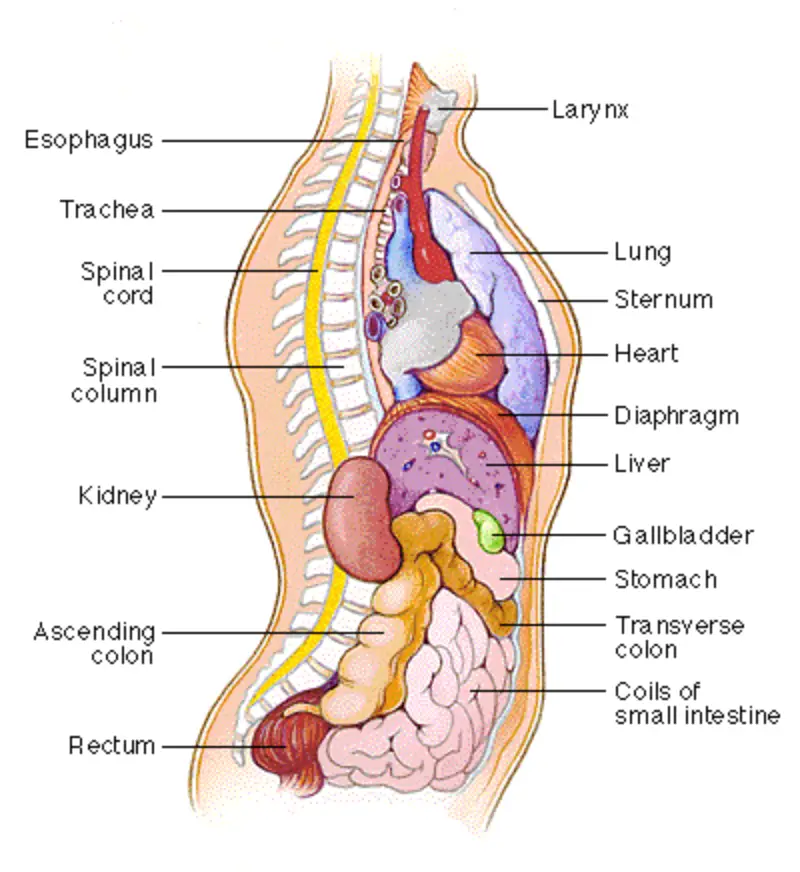
Organs of the Chest (Thoracic Cavity)
The thoracic cavity is enclosed by the rib cage, sternum, and vertebrae. It contains the heart, lungs, esophagus, trachea, and major blood vessels such as the aorta and vena cava.
1. Heart
- Location: The heart is located slightly left of the midline in the mediastinum, between the lungs.
- Structure: The heart has four chambers (two atria and two ventricles) and is enclosed in a fibrous sac called the pericardium.
- Function: The heart pumps oxygenated blood to the body (systemic circulation) via the left side, and deoxygenated blood to the lungs (pulmonary circulation) via the right side. It is responsible for maintaining the circulation of blood through the entire body, delivering oxygen and nutrients to tissues while removing waste products.
- Clinical Relevance: Common heart disorders include coronary artery disease, heart attacks (myocardial infarction), arrhythmias, and heart failure. Diagnostic methods include ECG, echocardiography, and angiography.
2. Lungs
- Location: The lungs are located on either side of the heart in the pleural cavities.
- Structure: The right lung has three lobes (superior, middle, and inferior), while the left lung has two lobes (superior and inferior). The lungs contain millions of tiny air sacs called alveoli, where gas exchange occurs.
- Function: The lungs facilitate the exchange of oxygen and carbon dioxide between the air and the blood. Oxygen is absorbed into the bloodstream, and carbon dioxide, a metabolic waste product, is expelled from the body during exhalation.
- Clinical Relevance: Lung diseases include asthma, chronic obstructive pulmonary disease (COPD), pneumonia, lung cancer, and tuberculosis. Diagnostic methods include chest X-rays, spirometry, CT scans, and bronchoscopy.
3. Esophagus
- Location: The esophagus runs behind the trachea and heart and connects the pharynx (throat) to the stomach.
- Structure: It is a muscular tube that extends through the thoracic cavity, passing through the diaphragm to reach the stomach.
- Function: The esophagus transports food and liquids from the mouth to the stomach via peristalsis (a series of wave-like muscle contractions).
- Clinical Relevance: Conditions affecting the esophagus include gastroesophageal reflux disease (GERD), esophagitis, and esophageal cancer. Diagnosis is often done using endoscopy and barium swallow studies.
4. Trachea and Bronchi
- Location: The trachea (windpipe) is located in front of the esophagus and extends from the larynx to the lungs.
- Structure: The trachea divides into two main bronchi, which further branch into smaller bronchi and bronchioles within the lungs.
- Function: The trachea and bronchi conduct air to the lungs. The trachea is lined with cilia and mucus to trap particles and pathogens before they enter the lungs.
- Clinical Relevance: Disorders include tracheitis, bronchitis, and tracheal stenosis. Bronchoscopy and CT imaging are common diagnostic tools.
5. Major Blood Vessels (Aorta, Superior and Inferior Vena Cava)
- Aorta: The aorta is the largest artery in the body and arises from the left ventricle of the heart, carrying oxygenated blood to the systemic circulation.
- Superior and Inferior Vena Cava: These large veins return deoxygenated blood from the body to the right atrium of the heart.
Organs of the Abdomen (Abdominal Cavity)
The abdominal cavity is located below the diaphragm and houses most of the organs involved in digestion, metabolism, and waste excretion. The abdominal organs include the stomach, intestines, liver, pancreas, spleen, kidneys, and bladder.
1. Stomach
- Location: The stomach is located in the upper left quadrant of the abdomen, just below the diaphragm.
- Structure: It is a J-shaped organ with regions including the fundus, body, and pylorus. The stomach is lined with gastric glands that secrete acid and enzymes.
- Function: The stomach breaks down food mechanically and chemically, producing chyme (a semi-liquid mixture of partially digested food) which is passed into the small intestine.
- Clinical Relevance: Common stomach conditions include gastritis, peptic ulcers, and gastric cancer. Diagnostic methods include endoscopy and barium studies.
2. Small Intestine
- Location: The small intestine extends from the stomach to the large intestine, occupying the central portion of the abdomen.
- Structure: It is divided into three segments: duodenum, jejunum, and ileum. The inner lining contains villi and microvilli to maximize nutrient absorption.
- Function: The small intestine is responsible for the majority of nutrient absorption. Digestive enzymes from the pancreas and bile from the liver aid in breaking down fats, proteins, and carbohydrates.
- Clinical Relevance: Disorders of the small intestine include celiac disease, Crohn’s disease, and small bowel obstructions. Diagnosis often involves endoscopy and imaging studies.
3. Large Intestine (Colon)
- Location: The large intestine surrounds the small intestine and extends from the ileum to the rectum.
- Structure: It is divided into the cecum, ascending colon, transverse colon, descending colon, sigmoid colon, and rectum.
- Function: The large intestine absorbs water and electrolytes from indigestible food matter, forming solid waste (feces) for elimination.
- Clinical Relevance: Common conditions include diverticulitis, colorectal cancer, and irritable bowel syndrome (IBS). Colonoscopy is a key diagnostic tool.
4. Liver
- Location: The liver is located in the upper right quadrant of the abdomen, beneath the diaphragm.
- Structure: The liver is a large, reddish-brown organ divided into four lobes. It is connected to the gallbladder and intestines via the bile ducts.
- Function: The liver plays a key role in metabolism, detoxification, and nutrient storage. It produces bile to aid in fat digestion, processes nutrients from the digestive tract, and detoxifies harmful substances.
- Clinical Relevance: Liver diseases include hepatitis, cirrhosis, and liver cancer. Blood tests, ultrasound, and liver biopsy are common diagnostic methods.
5. Gallbladder
- Location: The gallbladder is a small, pear-shaped organ located beneath the liver.
- Structure: It stores and concentrates bile produced by the liver.
- Function: The gallbladder releases bile into the small intestine to assist in the digestion and absorption of fats.
- Clinical Relevance: Gallbladder diseases include cholelithiasis (gallstones) and cholecystitis (inflammation of the gallbladder). Diagnosis often involves ultrasound and cholecystography.
6. Pancreas
- Location: The pancreas is located behind the stomach and extends across the upper abdomen.
- Structure: It has both exocrine (digestive enzyme-producing) and endocrine (hormone-producing) functions.
- Function: The pancreas secretes digestive enzymes into the small intestine and produces hormones like insulin and glucagon, which regulate blood sugar levels.
- Clinical Relevance: Pancreatic disorders include pancreatitis, pancreatic cancer, and diabetes. Diagnostic methods include imaging studies, blood tests, and biopsy.
7. Spleen
- Location: The spleen is located in the upper left quadrant of the abdomen, near the stomach.
- Structure: It is a soft, purplish organ that is part of the lymphatic system.
- Function: The spleen filters and recycles old red blood cells, produces lymphocytes (immune cells), and stores blood platelets.
- Clinical Relevance: Conditions include splenomegaly (enlarged spleen), splenic rupture, and lymphoma. Imaging tests such as ultrasound and CT scans are used for diagnosis.
8. Kidneys
- Location: The kidneys are located on either side of the spine, just below the ribcage in the retroperitoneal space.
- Structure: The kidneys are bean-shaped organs with an outer cortex and inner medulla, containing structures called nephrons that filter the blood.
- Function:
The kidneys filter waste products from the blood to form urine, regulate electrolytes and blood pressure, and maintain fluid balance.
- Clinical Relevance: Kidney disorders include chronic kidney disease, kidney stones, and renal failure. Diagnostic tools include blood tests, urinalysis, and imaging studies like ultrasound and CT scans.
9. Urinary Bladder
- Location: The bladder is located in the lower abdomen, behind the pubic bone.
- Structure: It is a hollow, muscular organ that stores urine before it is excreted from the body.
- Function: The bladder temporarily stores urine, which is produced by the kidneys and passed down through the ureters. When full, it contracts to expel urine through the urethra.
- Clinical Relevance: Bladder disorders include urinary incontinence, bladder infections (cystitis), and bladder cancer. Diagnosis is often performed using urinalysis, ultrasound, and cystoscopy.
Clinical Relevance of Abdominal and Thoracic Organs
Diseases and disorders affecting the chest and abdominal organs can have significant clinical consequences, impacting respiratory function, digestion, circulation, and metabolic processes. Early detection and treatment are essential for managing conditions related to these vital organs. Diagnostic tools such as imaging studies (ultrasound, CT scans, MRIs), endoscopic procedures, and blood tests are widely used in the diagnosis and management of chest and abdominal diseases.
Conclusion
The organs of the chest and abdomen play critical roles in maintaining vital physiological functions, from breathing and circulation to digestion and excretion. A thorough understanding of the anatomy and function of these organs is essential for the diagnosis and treatment of many common and life-threatening conditions. Advances in medical imaging and minimally invasive procedures continue to improve patient outcomes and provide better insights into the complexities of thoracic and abdominal diseases.
References
- Standring, S. (2020). Gray’s Anatomy: The Anatomical Basis of Clinical Practice (42nd ed.). Elsevier.
- Moore, K. L., Dalley, A. F., & Agur, A. M. R. (2013). Clinically Oriented Anatomy (7th ed.). Lippincott Williams & Wilkins.
- Netter, F. H. (2014). Atlas of Human Anatomy (6th ed.). Elsevier.
- Fauci, A. S., Braunwald, E., Kasper, D. L., Hauser, S. L., & Longo, D. L. (2008). Harrison’s Principles of Internal Medicine (17th ed.). McGraw-Hill.
This comprehensive overview highlights the structure, function, and clinical relevance of the organs in the chest and abdomen, underscoring their importance to overall health and survival.
🎓 Want to become a certified instructor?
This lesson is part of our FREE Anatomy course. Create a free account to track your progress and earn your certificate!