The Bones of the Lower Limb: Structure, Function, and Clinical Relevance
Abstract
The lower limb, consisting of the hip, thigh, leg, and foot, plays a crucial role in locomotion, weight-bearing, and maintaining balance. It includes several bones: the femur, patella, tibia, fibula, and the bones of the foot. This article provides an in-depth examination of the anatomy, development, functions, and clinical significance of the bones of the lower limb.
Introduction
The bones of the lower limb are essential for supporting the body’s weight and enabling a wide range of movements necessary for walking, running, and other activities. These bones are robust and intricately connected to provide strength, stability, and flexibility. Understanding their anatomy and functions is fundamental in fields such as orthopedics, sports medicine, and physical therapy.
Anatomical Structure
The lower limb is divided into four main regions: the hip, thigh, leg, and foot. Each region contains specific bones with unique features and functions.
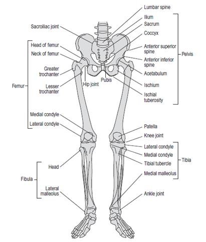
Hip
The hip includes the pelvic girdle, specifically the hip bone (os coxae), which connects the lower limb to the axial skeleton.
Hip Bone (Os Coxae)
The hip bone is formed by the fusion of three bones:
- Ilium: The broad, flaring portion of the hip bone.
- Ischium: The lower, posterior part.
- Pubis: The anterior part.
Key features include:
- Acetabulum: The socket that articulates with the head of the femur.
- Iliac Crest: The superior border of the ilium.
- Ischial Tuberosity: The part of the hip bone we sit on.
- Pubic Symphysis: The cartilaginous joint uniting the left and right pubic bones.
Thigh
The thigh contains one major bone:
- Femur (Thigh Bone)
Femur
The femur is the longest and strongest bone in the body. Key anatomical features include:
- Head: Articulates with the acetabulum of the hip bone.
- Neck: The narrow region just below the head.
- Greater and Lesser Trochanters: Bony prominences for muscle attachment.
- Shaft: The long, cylindrical portion.
- Medial and Lateral Condyles: Articulate with the tibia and patella at the knee joint.
Leg
The leg comprises two parallel bones:
- Tibia (Shin Bone)
- Fibula
Tibia
The tibia is the larger and more medial of the two leg bones. Important features include:
- Medial and Lateral Condyles: Articulate with the femur.
- Tibial Tuberosity: The site of attachment for the patellar ligament.
- Medial Malleolus: The bony prominence on the inner side of the ankle.
Fibula
The fibula is the thinner, lateral bone of the leg. Notable features include:
- Head: Articulates with the tibia.
- Lateral Malleolus: The bony prominence on the outer side of the ankle.
Foot
The foot consists of the tarsus, metatarsus, and phalanges. The bones of the foot include:
- Tarsals (Ankle Bones)
- Metatarsals (Foot Bones)
- Phalanges (Toe Bones)
Tarsals
The tarsus comprises seven bones:
- Calcaneus: The heel bone.
- Talus: Articulates with the tibia and fibula.
- Navicular, Cuboid, and Cuneiforms (Medial, Intermediate, Lateral): Contribute to the arch of the foot.
Metatarsals
The metatarsus consists of five metatarsal bones, numbered I to V from the medial to lateral side of the foot. Each metatarsal has a base, shaft, and head.
Phalanges
The phalanges are the bones of the toes. Each toe has three phalanges (proximal, middle, distal), except the big toe, which has two (proximal, distal).
Development
The bones of the lower limb develop through endochondral ossification:
- Mesenchymal Condensation: Formation of cartilaginous models during fetal development.
- Primary Ossification Centers: Develop in the diaphysis (shaft) of long bones.
- Secondary Ossification Centers: Appear in the epiphyses (ends) of long bones during childhood and adolescence, contributing to growth in length.
Function
The bones of the lower limb serve several vital functions:
- Support: Provide a framework for the body and support its weight during standing and movement.
- Movement: Facilitate a wide range of motions, including walking, running, jumping, and balance.
- Protection: Shield vital structures such as blood vessels, nerves, and muscles.
- Leverage: Act as levers to enhance the force and speed of movements.
Clinical Relevance
The bones of the lower limb are susceptible to various conditions and injuries:
- Fractures: Common in the femur, tibia, and fibula, often resulting from trauma or falls.
- Osteoarthritis: Degenerative joint disease affecting the hip, knee, and ankle joints.
- Osteoporosis: A condition that weakens bones, making them more prone to fractures.
- Ligament Injuries: Tears or sprains, particularly in the knee (e.g., ACL injuries).
- Plantar Fasciitis: Inflammation of the plantar fascia causing heel pain.
- Bunions: Deformities of the big toe joint.
Diagnostic and Therapeutic Approaches
Diagnosis of lower limb disorders typically involves clinical examination and imaging techniques such as X-rays, MRI, and CT scans. Treatment options vary depending on the condition and may include:
- Physical Therapy: Exercises to improve strength, flexibility, and function.
- Medications: Pain relievers, anti-inflammatory drugs, and bisphosphonates for osteoporosis.
- Surgical Interventions: Procedures such as fracture fixation, joint replacement, and ligament reconstruction.
- Orthotics and Braces: Devices to support and protect affected areas during healing.
Conclusion
The bones of the lower limb are essential for supporting the body, enabling movement, and maintaining balance. Understanding their anatomy, development, and associated pathologies is crucial for effective medical care. Advances in diagnostic techniques and treatment options continue to improve the management of lower limb disorders, enhancing patient outcomes.
References
- Standring, S. (2020). Gray’s Anatomy: The Anatomical Basis of Clinical Practice (42nd ed.). Elsevier.
- Moore, K. L., Dalley, A. F., & Agur, A. M. R. (2013). Clinically Oriented Anatomy (7th ed.). Lippincott Williams & Wilkins.
- Sadler, T. W. (2018). Langman’s Medical Embryology (14th ed.). Wolters Kluwer.
- Netter, F. H. (2014). Atlas of Human Anatomy (6th ed.). Elsevier.
- Rockwood, C. A., Green, D. P., & Bucholz, R. W. (2010). Rockwood and Green’s Fractures in Adults (7th ed.). Lippincott Williams & Wilkins.
This comprehensive examination of the bones of the lower limb highlights their complexity and importance, emphasizing the need for ongoing research and education in musculoskeletal health and disease management.
🎓 Want to become a certified instructor?
This lesson is part of our FREE Anatomy course. Create a free account to track your progress and earn your certificate!