The Muscles of the Head and Face: Structure, Function, and Clinical Relevance
The Muscles of the Head and Face: Structure, Function, and Clinical Relevance
Abstract
The muscles of the head and face are a complex group of muscles responsible for a wide range of functions, including facial expression, mastication, eye movement, and speech. These muscles are integral to communication, sensory functions, and the overall aesthetics of the face. This article explores the detailed anatomy, development, functions, and clinical significance of the muscles of the head and face.
Introduction
The muscles of the head and face are essential for daily activities, from expressing emotions to chewing food and speaking. These muscles can be divided into several groups based on their functions: facial expression, mastication, eye movement, and other specialized functions. Understanding the anatomy and function of these muscles is crucial in fields such as neurology, dentistry, plastic surgery, and speech therapy.
Anatomical Structure
The muscles of the head and face can be broadly categorized into four main groups:
- Muscles of Facial Expression
- Muscles of Mastication
- Extraocular Muscles
- Other Specialized Muscles
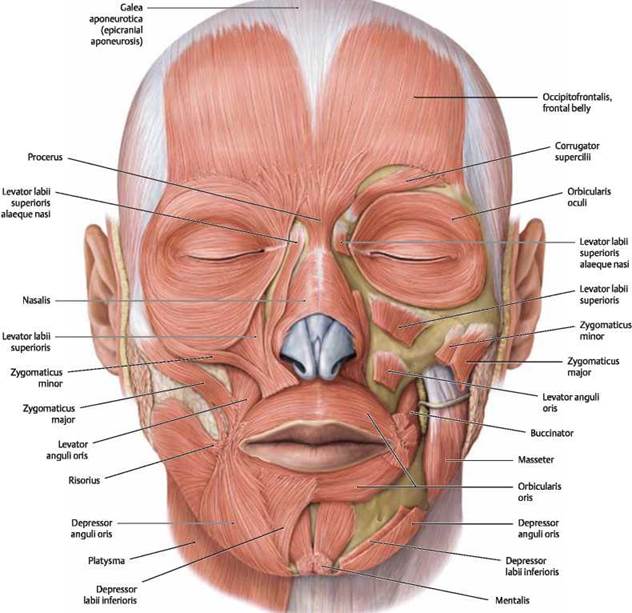
Muscles of Facial Expression
The muscles of facial expression are primarily responsible for moving the skin and soft tissues of the face to convey emotions. These muscles are innervated by the facial nerve (cranial nerve VII) and are unique in that they attach to the skin rather than bones. Key muscles include:
- Frontalis: Raises the eyebrows and wrinkles the forehead.
- Orbicularis Oculi: Encircles the eye, allowing for blinking and closing the eyelids.
- Orbicularis Oris: Encircles the mouth, enabling the lips to close and pucker (e.g., in kissing or whistling).
- Zygomaticus Major and Minor: Elevate the corners of the mouth, producing a smile.
- Buccinator: Compresses the cheek, aiding in chewing and blowing.
- Depressor Anguli Oris: Lowers the corners of the mouth, producing a frown.
- Platysma: Tenses the skin of the neck and depresses the mandible.
Muscles of Mastication
The muscles of mastication are responsible for the movement of the jaw during chewing and speaking. These muscles are innervated by the mandibular branch of the trigeminal nerve (cranial nerve V3). The four primary muscles of mastication are:
- Masseter: Elevates the mandible, closing the jaw.
- Temporalis: Elevates and retracts the mandible.
- Medial Pterygoid: Elevates the mandible and assists in grinding movements.
- Lateral Pterygoid: Protracts the mandible and allows for side-to-side grinding movements.
Extraocular Muscles
The extraocular muscles control the movement of the eyeball and the upper eyelid. These muscles are crucial for precise eye movements and are innervated by three cranial nerves: oculomotor (cranial nerve III), trochlear (cranial nerve IV), and abducens (cranial nerve VI). The six extraocular muscles are:
- Superior Rectus: Elevates the eye.
- Inferior Rectus: Depresses the eye.
- Medial Rectus: Adducts the eye.
- Lateral Rectus: Abducts the eye.
- Superior Oblique: Depresses and intorts the eye.
- Inferior Oblique: Elevates and extorts the eye.
Other Specialized Muscles
There are several other specialized muscles in the head and face that perform unique functions:
- Levator Palpebrae Superioris: Elevates the upper eyelid, innervated by the oculomotor nerve (cranial nerve III).
- Digastric, Mylohyoid, and Geniohyoid: These suprahyoid muscles assist in elevating the hyoid bone and larynx during swallowing and speech.
- Muscles of the Tongue: The intrinsic and extrinsic muscles of the tongue control its shape and movement, essential for speech, swallowing, and taste. These muscles are primarily innervated by the hypoglossal nerve (cranial nerve XII).
Development
The muscles of the head and face develop from the mesodermal layer of the embryo, with specific groups arising from different pharyngeal arches:
- First Pharyngeal Arch: Gives rise to the muscles of mastication.
- Second Pharyngeal Arch: Develops into the muscles of facial expression.
- Ocular Muscles: Develop from preotic myotomes, which are specific segments of the mesoderm in the head.
Function
The muscles of the head and face perform several critical functions:
- Facial Expression: Convey emotions and social signals through movement of the skin and facial structures.
- Mastication: Enable the grinding and chewing of food, crucial for digestion.
- Eye Movement: Allow for precise control of gaze and focus.
- Speech: Facilitate the articulation of sounds and words.
- Protection: Movements like blinking protect the eyes from injury and dehydration.
Clinical Relevance
Disorders and injuries affecting the muscles of the head and face can have significant clinical implications:
- Bell’s Palsy: A condition resulting in sudden weakness or paralysis of the muscles on one side of the face due to facial nerve dysfunction.
- Trigeminal Neuralgia: A chronic pain condition affecting the trigeminal nerve, causing intense facial pain.
- Temporomandibular Joint Disorder (TMD): Involves dysfunction and pain in the muscles of mastication and the temporomandibular joint.
- Strabismus: A condition where the eyes do not align properly due to dysfunction of the extraocular muscles.
- Myasthenia Gravis: An autoimmune disorder that causes weakness in the muscles, including those of the face, often affecting eye and facial movements.
- Facial Trauma: Injuries such as fractures or lacerations can damage facial muscles, leading to functional and aesthetic issues.
Diagnostic and Therapeutic Approaches
Diagnosis of disorders affecting the muscles of the head and face often involves clinical examination, imaging techniques (such as MRI or CT scans), and electromyography (EMG) to assess muscle function. Treatment varies depending on the condition and may include:
- Medications: Anti-inflammatory drugs, muscle relaxants, or antispasmodics.
- Physical Therapy: Exercises to strengthen or relax muscles, improve coordination, and alleviate pain.
- Botulinum Toxin Injections (Botox): Used to manage muscle spasticity or treat conditions like chronic migraine and TMD.
- Surgical Interventions: May be required for structural issues such as strabismus or facial nerve decompression in severe cases of Bell’s palsy.
- Speech Therapy: Essential for individuals with speech disorders related to facial muscle dysfunction.
Conclusion
The muscles of the head and face are vital for a wide range of functions, from basic survival activities like eating and speaking to complex social interactions through facial expressions. Understanding their anatomy, development, and potential pathologies is crucial for effective medical care. Advances in diagnostic and therapeutic techniques continue to improve the management of conditions affecting these muscles, enhancing the quality of life for patients.
References
- Standring, S. (2020). Gray’s Anatomy: The Anatomical Basis of Clinical Practice (42nd ed.). Elsevier.
- Moore, K. L., Dalley, A. F., & Agur, A. M. R. (2013). Clinically Oriented Anatomy (7th ed.). Lippincott Williams & Wilkins.
- Netter, F. H. (2014). Atlas of Human Anatomy (6th ed.). Elsevier.
- Williams, P. L., & Warwick, R. (1980). Gray’s Anatomy (36th ed.). Churchill Livingstone.
- Agur, A. M. R., & Dalley, A. F. (2017). Grant’s Atlas of Anatomy (14th ed.). Lippincott Williams & Wilkins.
- Lieberman, D. E., Hallgrimsson, B., & Parsons, T. E. (2008). Developmental Factors in the Evolution of the Primate Facial Skeleton. Journal of Anatomy, 212(4), 426-442.
This comprehensive exploration of the muscles of the head and face emphasizes their complexity and importance, highlighting the need for ongoing research and education in neuromuscular health and disease management.
🎓 Want to become a certified instructor?
This lesson is part of our FREE Anatomy course. Create a free account to track your progress and earn your certificate!